. 2025 Mar 24;26(1):67.
doi: 10.1186/s40360-025-00901-7.

Effects of adenosine triphosphate, thiamine pyrophosphate, melatonin, and liv-52 on subacute pyrazinamide proliferation hepatotoxicity in rats

Affiliations

Effects of adenosine triphosphate, thiamine pyrophosphate, melatonin, and liv-52 on subacute pyrazinamide proliferation hepatotoxicity in rats

Sedat Ciftel et al. BMC Pharmacol Toxicol. .

Abstract

Background: Hepatotoxicity of pyrazinamide, an antituberculosis drug, limits its therapeutic use and oxidative stress has been implicated in this toxicity. This study investigated the protective effects of adenosine triphosphate (ATP), thiamine pyrophosphate (TPP), melatonin, and Liv-52, which have previously been shown antioxidant activities, on pyrazinamide-induced hepatotoxicity.

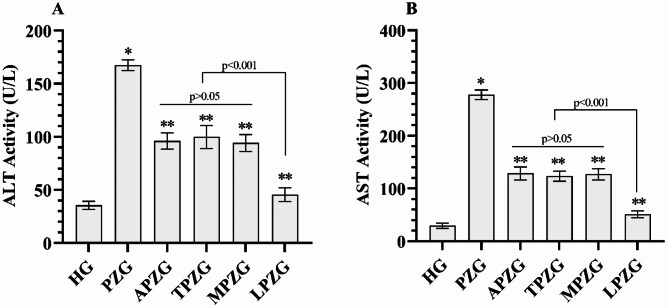
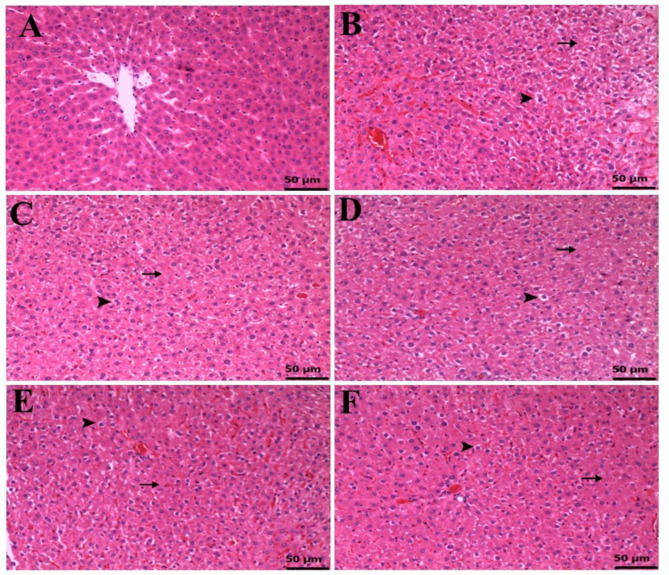
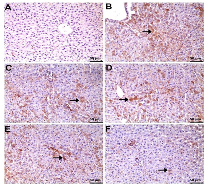
Methods: 36 albino Wistar male rats were divided into randomized six groups; healthy (HG), pyrazinamide (PZG), ATP + pyrazinamide (APZG), TPP + pyrazinamide (TPZG), melatonin + pyrazinamide (MPZG) and Liv-52 + pyrazinamide (LPZG) groups. ATP 4 mg/kg and TPP 25 mg/kg were administered intraperitoneally (IP). Melatonin 10 mg/kg and Liv-52 20 mg/kg were given orally. One hour after administration of ATP, TPP, melatonin, and Liv-52, 250 mg/kg pyrazinamide was applied orally to all rats except HG group. The treatment was repeated (1 × 1) for 4 weeks. Then, blood samples were taken for determination of alanine aminotransferase (ALT) and aspartate aminotransferase (AST) activities. Immediately after, the rats were euthanized with thiopental sodium (50 mg/kg, IP), and the livers were removed. The tissues were analyzed for malondialdehyde (MDA), total glutathione (tGSH), superoxide dismutase (SOD), and catalase (CAT) also hydropic degeneration, necrosis, and apoptosis (caspase 3) were examined.One-Way ANOVA was used in biochemical analyses and Tukey test was used as post-hoc. For histopathological and immunohistochemical analysis, the Kruskal-Wallis test was used and Dunn's test as a post-hoc.

Results: Pyrazinamide increased MDA land decreased tGSH, SOD, and CAT levels in liver tissues (p < 0.001). It also increased serum ALT and AST activities and caused severe hydropic degeneration and necrosis in liver tissue (p < 0.001). ATP, TPP, melatonin, and Liv-52 significantly prevented the biochemical and histopathological changes induced by pyrazinamide (p < 0.05). On the other hand, Liv-52 was more successful than other potential protectors in protecting liver tissue from pyrazinamide damage (p < 0.05).

Conclusions: ATP, TPP, melatonin, and Liv-52 can be used to protect liver tissue from pyrazinamide-induced hepatotoxicity in rats.

Keywords: ATP; Antioxidant effect; Liv-52; Liver toxicity; Pyrazinamide; Thiamine pyrophosphate.


Conflict of interest statement

Declarations. Ethics approval and consent to participate: The Erzincan Binali Yıldırım University, local Animal Experimentation Ethics Committee approved the procedures (Date: 29.08.2024, meeting no: 08/31). Consent for publication: Not applicable. Competing interests: The authors declare no competing interests.

Figures

Fig. 1
Fig. 1
(A-D) Evaluation of oxidant and antioxidant amounts in liver tissues of experimental groups. Bars shows mean ± dtandard deviation, n = 6. *; p < 0.001 vs. HG, **; p < 0.001 vs. PZG. MDA; malondialdehyde, tGSH; total glutathione, SOD; superoxide dismutase, CAT; catalase, HG; healthy group, PZG; pyrazinamide group, APZG; adenosine triphosphate + pyrazinamide group, TPZG; thiamine pyrophosphate + pyrazinamide group, MPZG; melatonin + pyrazinamide group, LPZG; Liv-52 + pyrazinamide group
Fig. 2
Fig. 2
(A-B) Evaluation of liver function tests in the serum of the experimental groups. Bars shows mean ± dtandard deviation, n = 6. *; p < 0.001 vs. HG, **; p < 0.001 vs. PZG. ALT; alanine aminotransferase, AST; aspartate aminotransferase, HG; healthy group, PZG; pyrazinamide group, APZG; adenosine triphosphate + pyrazinamide group, TPZG; thiamine pyrophosphate + pyrazinamide group, MPZG; melatonin + pyrazinamide group, LPZG; Liv-52 + pyrazinamide group
Fig. 3
Fig. 3
(A-F) Histological appearances of liver tissues of HG (A), PZG (B), APZG (C), TPZG (D), MPZG (E), and LPZG (F) groups (Hematoxylin-eosin staining). A: Normal histological appearance of liver tissue. B: Severe hydropic degeneration (arrowhead) and necrosis (arrow) in liver tissue. HG; healthy group, PZG; pyrazinamide group. C: Moderate hydropic degeneration (arrowhead) and necrosis (arrow) appearance in liver tissue. D: Moderate hydropic degeneration (arrowhead) and necrosis (arrow) appearance in liver tissue. E: Moderate hydropic degeneration (arrowhead) and necrosis (arrow) appearance in liver tissue. F: Mild hydropic degeneration (arrowhead) and necrosis (arrow) appearance in liver tissue. HG; healthy group, PZG; pyrazinamide group, APZG; adenosine triphosphate + pyrazinamide group, TPZG; thiamine pyrophosphate + pyrazinamide group, MPZG; melatonin + pyrazinamide group, LPZG; Liv-52 + pyrazinamide group
Fig. 4
Fig. 4
(A-F) Caspase 3 immunopositivity in liver tissue of HG (A), PZG (B), ATPG (C), TPZG (D), MPZG (E), and LPZG (F) groups (Immunohistochemistry). A: Immun negativity B: Severe immunopositivity (arrow). C: Moderate immunopositivity (arrow). D: Moderate immunopositivity (arrow). E: Moderate immunopositivity (arrow). F: Mild immunopositivity (arrow). HG; healthy group, PZG; pyrazinamide group, APZG; adenosine triphosphate + pyrazinamide group, TPZG; thiamine pyrophosphate + pyrazinamide group, MPZG; melatonin + pyrazinamide group, LPZG; Liv-52 + pyrazinamide group

Similar articles

References

    1. Hussain Z, Zhu J, Ma X. Metabolism and hepatotoxicity of pyrazinamide, an antituberculosis drug. Drug Metab Dispos. 2021;49:679–82. - PMC - PubMed
    1. Gopal P, Grüber G, Dartois V, Dick T. Pharmacological and molecular mechanisms behind the sterilizing activity of pyrazinamide. Trends Pharmacol Sci. 2019;40:930–40. - PMC - PubMed
    1. Lu P, Haagsma AC, Pham H, Maaskant JJ, Mol S, Lill H, Bald D. Pyrazinoic acid decreases the proton motive force, respiratory ATP synthesis activity, and cellular ATP levels. Antimicrob Agents Chemother. 2011;55:5354–7. - PMC - PubMed
    1. Krug S, Gupta M, Kumar P, et al. Inhibition of host PARP1 contributes to the anti-inflammatory and antitubercular activity of pyrazinamide. Nat Commun. 2023;14:8161. - PMC - PubMed
    1. Wang YC, Chen KH, Chen YL, et al. Pyrazinamide related prolonged drug-induced liver injury: A case report. Med (Baltim). 2022;101:e30955. - PMC - PubMed

MeSH terms