

Added value of radiological staging to clinical examination in different histopathological subtypes of uterine cervical cancer: A retrospective study
- PMID: 40129448
- PMCID: PMC11930720
- DOI: 10.1016/j.eurox.2025.100376
Added value of radiological staging to clinical examination in different histopathological subtypes of uterine cervical cancer: A retrospective study
Abstract
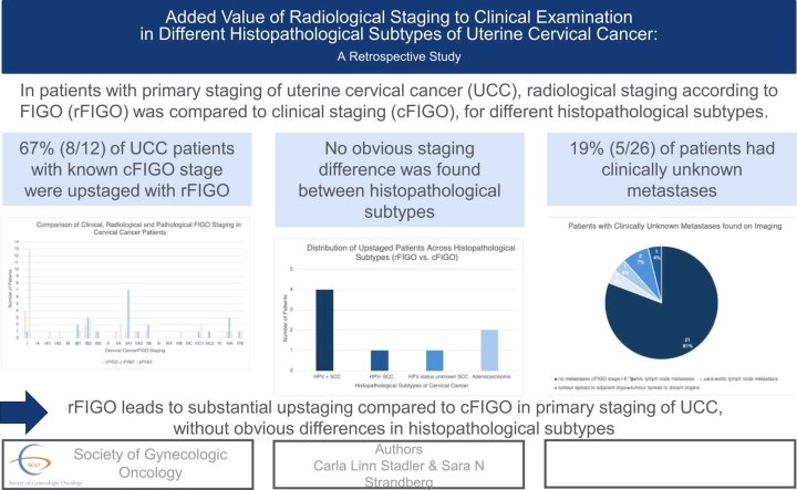
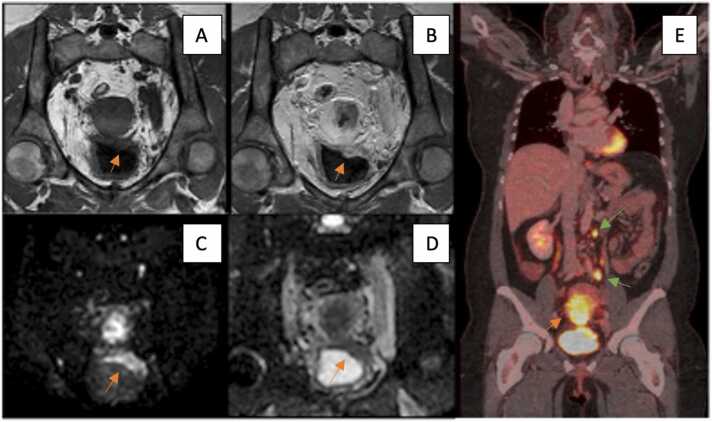
Objective: Accurate staging of uterine cervical cancer (UCC) is crucial for treatment guidance and prognostic predictions. This study investigated the added value of conventional diagnostic imaging for different histopathological subtypes of UCC by comparing clinical staging according to International Federation of Gynaecology and Obstetrics staging system (cFIGO) and radiological staging (rFIGO) with histopathological staging (pFIGO) as reference.
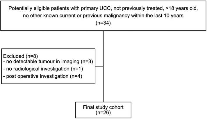
Methods: 26 consecutive patients with UCC from the retrospective part of the PRODIGYN study (ethical approval number 2022-04207-01; NCT05855941) were included in the present study. Data from study participants was collected from radiological and histopathological records 2016-2022 at the University hospital of Umeå. Staging was assessed according to the FIGO 2018 staging system. Statistical analysis included descriptive statistics and Cohen's weighted kappa coefficient (κ) for calculation of agreement between cFIGO and rFIGO, and between rFIGO and pFIGO.
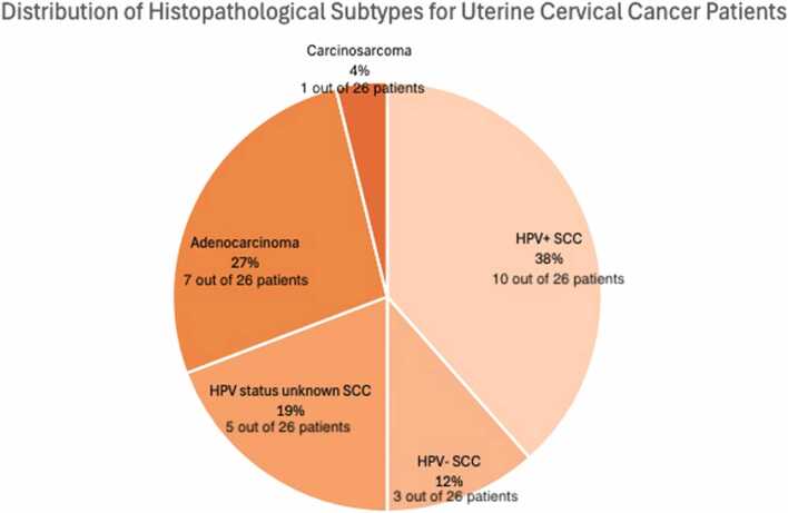
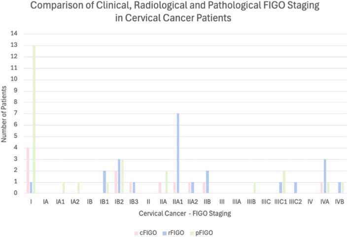
Results: With rFIGO staging, more advanced disease stages were found in 67 % (8/12 patients with known cFIGO). Poor agreement was found between cFIGO and rFIGO (κ =0.057) and between rFIGO and pFIGO (κ= 0169). Among the patients with squamous cell carcinoma (SCC) positive for human papilloma virus (HPV+), 67 % (4/6) were assigned a higher stage by rFIGO compared to cFIGO. For the single patients with HPV-negative SCC and HPV status unknown SCC, both were upstaged by rFIGO. In the case of adenocarcinomas, 67 % (2/3) of the patients were assigned a higher stage with rFIGO.
Conclusions: In primary staging of UCC, rFIGO leads to substantial up-staging compared to cFIGO, without obvious differences in subtypes.
Keywords: Adenocarcinoma; Computed Tomography; Magnetic Resonance Imaging; Positron Emission Tomography Computed Tomography; Squamous Cell Neoplasms; Uterine cervical neoplasms.
© 2025 The Authors.
Conflict of interest statement
The authors declare that they have no known competing financial interests or personal relationships that could have appeared to influence the work reported in this paper.
Figures
References
-
- Cancer Today. Accessed April 16, 2024. 〈https://gco.iarc.fr/today/en〉.
-
- Cervical Cancer - StatPearls - NCBI Bookshelf. Accessed April 24, 2024. 〈https://www.ncbi.nlm.nih.gov/books/NBK431093/〉.
-
- II. Complementary data on cervical cancer prevention. Accessed April 17, 2024. 〈www.hpvcentre.net〉.
LinkOut - more resources
Full Text Sources
Research Materials
