Case Reports
. 2025 Mar 10:11:1463428.
doi: 10.3389/fvets.2024.1463428. eCollection 2024.

Case report: Endolymphatic system disease in elasmobranchs: clinical presentation, diagnosis, and treatment strategies

Affiliations
Case Reports

Case report: Endolymphatic system disease in elasmobranchs: clinical presentation, diagnosis, and treatment strategies

Whitney Greene et al. Front Vet Sci. .

Abstract

The inner ear is an often overlooked system in elasmobranchs with few documented reports of disease or other abnormalities in the literature. Similar to terrestrial vertebrates, it is located in the cranium, and there are multiple components to the ear of elasmobranchs including a pair of membranous labyrinths each with three semicircular canals and four chambers or end organs (the saccule, the lagena, the utricle and the macula neglecta) making up the endolymphatic system (ELS). There is species variability among the inner ear anatomy of elasmobranchs, and this may play a role in disease development, progression, and treatment outcomes. Also similar to terrestrial vertebrates, this system plays a key role in hearing, acceleration, and orientation. When affected, clinical signs may include localized areas of swelling or stoma development along the dorsal midline of the head at the endolymphatic pores, atypical swimming behaviors consistent with vestibular disease (spiraling/spinning or barrel rolling, or tilting to one side), and anorexia. Less frequently, the eyes may also be affected and present with exophthalmia, hyphema, and/or panophthalmitis. Herein are case series from five institutions representing a variety of elasmobranch species affected with ELS disease with discussion of anatomy, clinical presentation, diagnostics, etiology, treatment, and outcomes. Endolymphatic disease may be clinically underdiagnosed in elasmobranchs and mistaken for other diseases such as superficial subcutaneous or subdermal abscesses, focal dermatitis, or neuropathies presumed to not be associated with the inner ear system. In addition, disease may be occult for a long period of time prior to overt manifestation of signs or chronic with waxing and waning clinical signs, likely because of anatomy and resultant treatment challenges. Awareness and additional research may help to promote timely identification, improve diagnostic and treatment options, and help to optimize individual animal welfare.

Keywords: endolymphatic pore; inner ear; labyrinthitis; neurologic disease; neuropathy; otic disease; shark; stingray.


Conflict of interest statement

RD-V and NM were employed by The Walt Disney Company. NP was employed by Sea Life Park Hawaii. The remaining authors declare that the research was conducted in the absence of any commercial or financial relationships that could be construed as a potential conflict of interest.

Figures

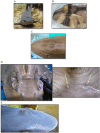
Figure 1
Figure 1
Endolymphatic pores in normal elasmobranchs. (A) Bamboo shark (Chiloscyllium punctatum), close up. (B) Zebra shark (Stegostoma tigrinum) post-mortem cerebrospinal fluid collection. (C) Bowmouth guitarfish (Rhina ancylostoma). (D) Chilean ray (Urotrygon chilensis). (E) Coral catshark (Atelomycterus marmoratus). (F) Spotted eagle ray (Aetobatus narinari) cerebrospinal fluid collection, post-mortem.
Figure 2
Figure 2
Line drawing of basic inner ear of elasmobranchs, representing general anatomy. This is a representation, differences may include variations in size/depth of the parietal fossa, in the endolymphatic duct’s direction and width/volume and shape of the perilymph bathing the labyrinth within the chondrocranium and the size of the end organs. Adapted from Chang and Okihiro (5).
Figure 3
Figure 3
Swelling along the dorsal midline associated with endolymphatic system disease in various elasmobranch species. (A) White tip reef shark (Triaenodon obesus); (B) Port Jackson shark (Heterodontus portusjacksoni); (C) Bamboo shark (Chiloscyllium punctatum); (D) Bowmouth guitarfish (Rhina ancylostoma), zoomed out and up close; (E) White tip reef shark (Triaenodon obesus).
Figure 4
Figure 4
Various drains and debridement as part of a multimodal treatment plan for endolymphatic disease in elasmobranch species. (A) Porcupine Ray (Urogymnus asperrimus); (B) Bowmouth guitarfish (Rhina ancylostoma) with flushing; (C) White tip reef shark (Triaenodon obesus) tagging post aspiration.
Figure 5
Figure 5
Pre and post contrast radiographic images of a porcupine ray (Urogymnus asperrimus) with endolymphatic system disease. (A) Survey view, ventrodorsal (VD) view and lateral. (B) Post iodinated-contrast infused into the affected area, VD view and lateral.
Figure 6
Figure 6
Surgery and catheterization for treatment of endolymphatic disease in a Bowmouth guitarfish (Rhina ancylostoma). (A) Clinical presentation; (B) Flushing the parietal fossa during first exam (Procedure #1); (C) Catheterization into parietal fossa during procedure #2; (D) Surgical approach to fossa and catheterization into perilymphatic fenestration during procedure #3; (E) Closure of surgical site and securing catheter; (F) View of catheterization at necropsy with a metal probe inserted into the endolymphatic pore, up close and yellow arrows indicate the endolymphatic pore.
Figure 7
Figure 7
Postmortem examples of endolymphatic disease in elasmobranchs. (A) Cranial to caudal cross sections of the inner ear at necropsy in a bowmouth guitarfish (Rhina ancylostoma); (B) The opening that was catheterized in a bowmouth guitarfish (Rhina ancylostoma) #2 from Case Series 1 with the probe is showing the direction of the fenestration into the otic capsule. (C) Necropsy of a Port Jackson shark (Heterodontus portusjacksoni) with unilateral labyrinthitis. The infected labyrinth (green arrows) shows severe congestion of the semicircular canals. (D) Adenocarcinoma in a whitetip reef shark (Triaenodon obesus) at necropsy.
Figure 8
Figure 8
Magnetic Resonance Imaging from the 3D-CISS sequence of the endolymphatic system in Port Jackson sharks (Heterodontus portusjacksoni). These images illustrate the absence of signal in the infected left membranous labyrinth. (A) Images highlighting the MR signal of the unaffected right membranous labyrinth and its corresponding 3D reconstruction. (B–D) 3D reconstruction of the head with successive transparency applied for virtual dissection of tissues to highlight the presence of the membranous labyrinth in the unaffected right inner ear, filled with lymph (green arrows).
Figure 9
Figure 9
Examples of various diagnostics for endolymphatic disease in elasmobranchs. (A) Marking of affected area for sample collection and treatment administration in a yellow ray (Urobatis jamaicensis); (B) Flushing of affected region as part of debridement in a white-spotted bamboo shark (Chiloscyllium plagiosum); (C) Flushing the affected region as part of debridement in a yellow ray (Urobatis jamaicensis); (D) Abscess aspiration in a porcupine ray (Urogymnus asperrimus); (E) Cerebrospinal fluid (CSF) collection in a bowmouth guitarfish (Rhina ancylostoma); (F) Cone-beam computed tomography of an anesthetized yellow ray (Urobatis jamaicensis), ventilated while raised out of water to maximize image quality.
Figure 10
Figure 10
Examples of cone beam CT (CBCT) images of right-sided otitis media in a bowmouth guitarfish (Rhina ancylostoma). (A) Transverse plane and (B) dorsal plane image demonstrating the expansile appearance and thinning of the mineral margins of the otic capsule. (C) Volume rendered image of the same individual viewed from dorsal with the endolymphatic duct segmented in green.

Similar articles

References

    1. Pereira N, Nunes GD, Baylina N, Peleteiro MC, Rosado L. Fusarium solani infection in the eye, brain, otic capsule and endolymphatic tubes in captive port Jackson sharks (Heterodontus portusjacksoni). In: Proceedings of the 33rd annual conference of the International Association for Aquatic Animal Medicine; (2002). p. 54-6.
    1. Pereira N, Faustino R, Faísca P, Veríssimo C, David H, Baylina N. Twenty years later: a new case of endolymphatic tube infection, internal otitis and meningitis caused by Fusarium Solani in a port Jackson shark (Heterodontus Portusjacksoni). In: Proceedings of the International Association for Aquatic Animal Medicine (IAAAM) meeting and conference (2018).
    1. Rich AF, Zendri F, Thornton S, Verin R. Meningoencephalitis with chondrocranial subcutaneous abscessation and septicaemia in a captive lemon shark (Negaprion brevirostris). J Fish Dis. (2021) 44:113–7. doi: 10.1111/jfd.13276 - DOI - PubMed
    1. Steele LM, Okihiro MS, Berlemont R, Dillon JG, Young KA, Hesami S, et al. . Carnobacterium maltaromaticum associated with meningoencephalitis and otitis in stranded common thresher sharks (Alopias vulpinus). Vet Pathol. (2022) 59:850–9. doi: 10.1177/03009858221102600, PMID: - DOI - PubMed
    1. Chang RK, Okihiro MS. A practical guide to necropsy of the elasmobranch chondrocranium and causes of mortality in wild and aquarium-housed California elasmobranchs. Front Vet Sci. (2024) 11:1410332. doi: 10.3389/fvets.2024.1410332, PMID: - DOI - PMC - PubMed

Publication types

LinkOut - more resources