. 2025 Mar 20:21:139-152.
doi: 10.2147/VHRM.S487327. eCollection 2025.

AQP1 Affects Necroptosis by Targeting RIPK1 in Endothelial Cells of Atherosclerosis

Affiliations

AQP1 Affects Necroptosis by Targeting RIPK1 in Endothelial Cells of Atherosclerosis

Ping Wang et al. Vasc Health Risk Manag. .

Abstract

Purpose: Aquaporin 1 (AQP1), a transmembrane water channel protein, has been implicated in the regulation of necroptosis. However, its specific role in atherosclerotic plaque stability through the modulation of necroptosis remains unclear. Therefore, in this study, we aim to investigate whether AQP1 influences necroptosis in atherosclerosis by binding to receptor-interacting serine/threonine-protein kinase 1 (RIPK1) and decreasing the expression of receptor-interacting serine/threonine-protein kinase 3 (RIPK3) and mixed lineage kinase domain-like pseudokinase (MLKL).

Patients and methods: The gene expression of AQP1 and necroptosis-associated genes significantly differ between atherosclerosis and normal groups. Genes linked to necroptosis were screened to influence the AS identified by weighted gene coexpression network analysis (WGCNA). Then we collected femoral atherosclerosis and normal aortic samples, further conducted single-cell sequencing and spatial transcriptomic methods to confirm the potential function and pathway of AQP1 in endothelial cells. Meanwhile, we overexpressed AQP1 in ox-LDL-treated endothelial cells in vitro.

Results: Firstly, via single-sample Gene Set Enrichment Analysis (ssGSEA) scores, we found that necroptosis plays the most important role among all ways of programmed cell death in two kinds of atherosclerosis. AQP1, RIPK1, RIPK3 and MLKL express differently in normal and atherosclerosis tissue by differentially expressed gene (DEG) analysis and Western Blot (WB). WGCNA analysis indicates that AQP1, MLKL and RIPK3 were significantly related to the AS. The area under the curve of the above hub genes was greater than 0.8 (AQP1 0.946, RIPK1 0.908, RIPK3 0.988, MLKL 0.863). We found AQP1 highly enriched in endothelial cells (ECs) by single-cell analysis. We sequenced the samples by spatial transcriptome and found that AQP1 was also mainly enriched in ECs both in expression and spatial location. With AQP1 overexpression in ECs, it significantly inhibited the expression of MLKL and RIPK3 and stimulated EC proliferation.

Conclusion: Our study identified that AQP1 suppresses atherosclerotic necroptosis by inhibiting the expression of RIPK3 and MLKL in ECs which might indicates that AQP1 plays a role in atherosclerosis. This new mechanism contributes to improving the diagnostic, prognostic, and therapeutic outcomes of atherosclerosis.

Keywords: carotid atherosclerosis; mechanism; necroptosis; plaque.


Conflict of interest statement

The authors declare no conflicts of interest in this work.

Figures

Figure 1
Figure 1
Results of transcriptome analysis of carotid and femoral artery samples. (a) The differentially expressed genes between normal and atherosclerosis samples including carotid and femoral samples are represented in a volcanic map. Necroptosis-related genes are labeled in the figure. (b) The results of immunoscores for normal and atherosclerosis samples including carotid and femoral samples are shown. The red items are the more incredible immune response in the atherosclerosis samples. The blue color was more common in normal samples.
Figure 2
Figure 2
Identification of genes associated with carotid and femoral atherosclerosis. (a) Volcano plot of the DEGs. Red, upregulation; blue, downregulation. (b) Heatmap of DEGs. (c-f) Box plots of AQP1, RIPK1, RIPK3 and MLKL in GSE100927 after data standardization. Blue represents disease sets, and red represents normal sets. (g) Heatmaps of AQP1, RIPK1, RIPK3 and MLKL among all samples into two groups. The blue color is the atherosclerotic sample group, and the red color is the normal group. G1, atherosclerosis group; G2, normal group. ****, P < 0.0001.
Figure 3
Figure 3
Identification of genes associated with carotid and femoral atherosclerosis. (a) Dendrogram based on a dissimilarity metric for all differentially expressed genes (1- TOM). (b) Relationship between module eigengenes and intraplaque bleeding as a heatmap. (c) ROC analysis of necroptosis genes. (d) Decreased AQP1 expression in carotid atherosclerosis tissues was detected by Western blotting. ****, P < 0.0001.
Figure 4
Figure 4
Procedures for sampling and analysis of carotid atherosclerotic plaques. The UMAP projection cluster scatter diagram was noted with clusters (a) and cell types (c). (b) The cell type and number of each sample are shown (GSM4837523, GSM4837524, GSM4837525, GSM4837526, GSM4837527, GSM4837528). (d) Heatmap shows the necroptosis genes distribution in each cell type. (e) shows the necroptosis genes (AQP1, RIPK1, RIPK3, MLKL) of each cell type and the distribution of these genes in the cluster map.
Figure 5
Figure 5
The single-cell analysis of femoral plaque and normal samples. (a) 20 clusters were noted with different colors in the dimplot based on UMAP projection. (b) shows the marker gene of each cell type. (c) The UMAP projection cluster scatter diagram was noted with cell type. Eight cell types were identified and labeled by matching marker. (d) is the cluster plot of normal and atherosclerosis cells respectively. (e) The expression level of AQP1, MLKL, RIPK1 and RIPK3 in each cluster. (f and g) HE staining of artery tissue of ST spots and unbiased clustering of ST spots in artery tissues. (h) cell types in single-cell analysis distribute in spatial position. (i) AQP1 expression level in whole spatial position. The darker the green color, the higher the expression. (j) plots displaying genes that are up-(red) or downregulated (blue) between normal and atherosclerosis groups for each cluster. Dashed lines denote fold change thresholds used when identifying DEGs.
Figure 6
Figure 6
(a and b) MTT proliferation assay and LDH release test in endothelial cells treated with different doses of ox-LDL for 48 h. (c and d) Western blot and qRT-PCR analysis of AQP1 expression following treatment of 300μg/mL ox-LDL for 48 h.
Figure 7
Figure 7
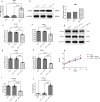
AQP1 overexpression affects the proliferation and apoptosis of endothelial cells and the expression of RIPK3 and MLKL. AQP1 expression after AQP1 overexpression by Western blot (a) and qRT-PCR (b). Changes of MLKL (c) and RIPK3 (d) before and after AQP1 knockdown in endothelial cells by qRT-PCR. Changes of MLKL (e) and RIPK3 (f) before and after AQP1 knockdown in endothelial cells by Western blot. (g) Western Blot gel images of MLKL and RIPK3. (h) Cell proliferation abilities were determined. (i) Cells undergoing apoptosis. (j) Colony formation ability.

Similar articles

References

    1. Skenteris NT, Hemme E, Delfos L, et al. Mast cells participate in smooth muscle cell reprogramming and atherosclerotic plaque calcification. Vascul Pharmacol. 2023;150:107167. doi:10.1016/j.vph.2023.107167 - DOI - PubMed
    1. Tsao CW, Aday AW, Almarzooq ZI, et al. Heart disease and stroke statistics-2023 update: a report from the American Heart Association. Circulation. 2023;147:e93–e621. doi:10.1161/cir.0000000000001123 - DOI - PubMed
    1. Puylaert P, Roth L, Van Praet M, et al. Effect of erythrophagocytosis-induced ferroptosis during angiogenesis in atherosclerotic plaques. Angiogenesis. 2023. doi:10.1007/s10456-023-09877-6 - DOI - PMC - PubMed
    1. Fidler TP, Xue C, Yalcinkaya M, et al. The AIM2 inflammasome exacerbates atherosclerosis in clonal haematopoiesis. Nature. 2021;592:296–301. doi:10.1038/s41586-021-03341-5 - DOI - PMC - PubMed
    1. Kluck GEG, Qian AS, Sakarya EH, Quach H, Deng YD, Trigatti BL. Apolipoprotein A1 protects against necrotic core development in atherosclerotic plaques: PDZK1-dependent high-density lipoprotein suppression of necroptosis in macrophages. Arterioscler Thromb Vasc Biol. 2023;43:45–63. doi:10.1161/ATVBAHA.122.318062 - DOI - PMC - PubMed

MeSH terms