. 2025 Feb 25;28(3):112099.
doi: 10.1016/j.isci.2025.112099. eCollection 2025 Mar 21.

Development of apical out trophoblast stem cell derived organoids to model early human pregnancy

Affiliations

Development of apical out trophoblast stem cell derived organoids to model early human pregnancy

J Zhou et al. iScience. .

Abstract

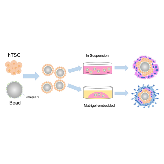
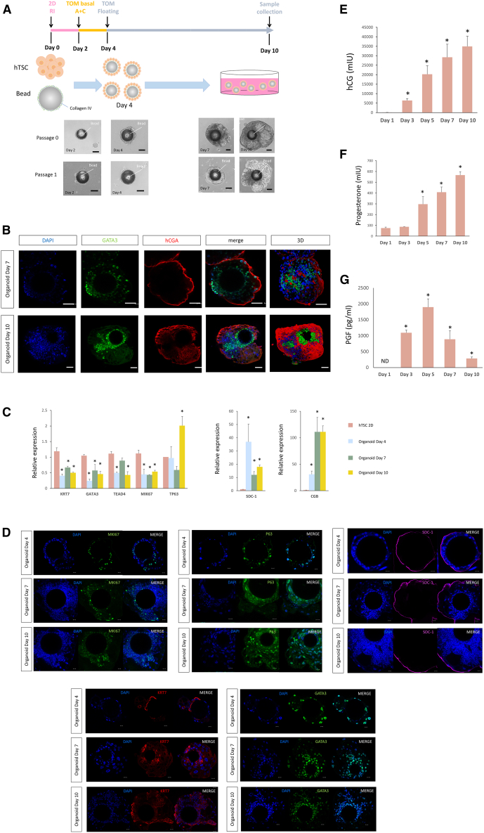
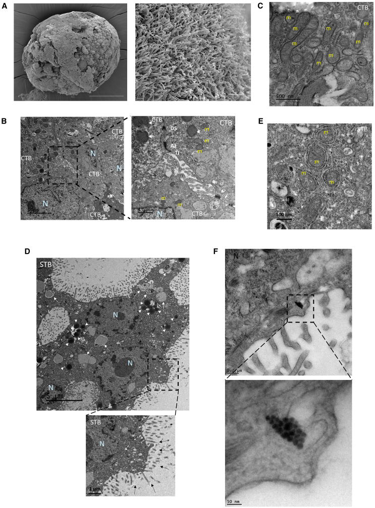
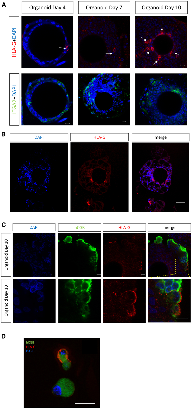
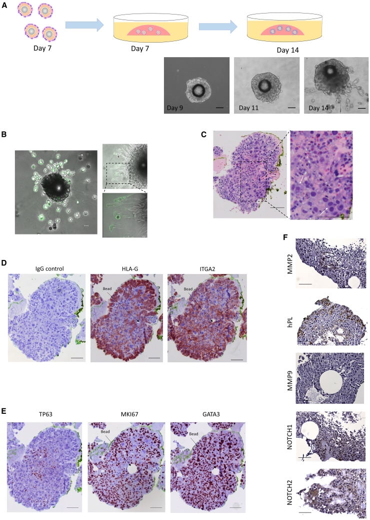
The development of trophoblast organoids has enabled investigation of placental physiology, disease, and early maternal-fetal interactions during a previously restricted stage of pregnancy. A key shortcoming in existing trophoblast organoid methodologies is the non-physiologic position of the syncytiotrophoblast (STB) within the inner portion of the organoid, which neither recapitulates in vivo placental villous morphology nor allows for facile modeling of STB exposure to the endometrium or the contents of the intervillous space. Here, we have successfully established apical-out human trophoblast stem cells (hTSC)-sourced organoids with STB forming on the surface of the organoid. These organoids can also be induced to give rise to the extravillous trophoblast (EVT) lineage, which invades into an extracellular matrix-based hydrogel. Compared to previous methods, our organoids more closely mimic developing human placental architecture, offering a novel platform to study normal and abnormal placental development and to model exposures to pharmaceuticals, pathogens, and environmental factors.

Keywords: Cell biology; Molecular biology; Physiology.


Conflict of interest statement

The authors declare that the research was conducted in the absence of any commercial or financial relationships that could be construed as a potential conflict of interest.

Figures

None
Graphical abstract
Figure 1
Figure 1
Establishment of apical-out trophoblast organoid cultures from hTSC cells (A) The hTSC cell line (CT27) was maintained under traditional 2D-culture conditions. To develop our organoid cultures, these cells were dispersed and incubated with collagen IV-coated beads in TSC medium supplemented with 5 μM Y27623 for 2 days (pink line). The medium was then switched to TOM basal medium containing A83-01(500 nM) and CHIR (1.5 μM) (TOM basal A + C) for an additional 2 days (yellow line). The developing organoids were then treated with complete TOM medium until day 10 (gray line). Bright-field images of hTSC-derived trophoblast organoids from passage 0 and passage 1 are depicted. Scale bars, 200μm. (B) Immunostaining of hCGA and GATA3 in STB organoids on Day 7 and Day 10. hCGA is expressed in multinucleated areas lining the outer surface of the organoids while the mononucleated cells closer to the center of the organoids are GATA3 positive. Reconstructed 3D confocal images showing the distribution of hCGA and GATA3. Scale bars, 50μm. (C) Quantitative real-time PCR analysis of CTB markers (upper panel) and STB markers (lower panel) in hTSC (2D culture) and STB organoids on Day 4, Day 7, and Day 10. KRT7, GATA3, TEAD4, and MKI67 transcript levels were higher in hTSC than in organoids, while hCGB and SDC1 transcript levels were much lower in hTSC than in organoids. Data were normalized to GAPDH expression. Presented values are the means ± SDs for three separate experiments Presented values are the means ± SDs for three separate experiments. Data followed over time were subjected to two-way ANOVA, followed by the Bonferroni test for pairwise comparisons. ∗p < 0.05. (D) Immunostaining for MKI67, GATA3, KRT7, P63, and SDC-1 in trophoblast organoids on Day 4, Day 7 and Day 10. Trophoblast cells in the center of the organoids express the CTB markers GATA3 and KRT7, MKI67 staining labeled proliferating trophoblast cell, and the STB marker SDC-1 is detected on the surface of the trophoblast organoids. Scale bars, 50μm. (E–G) Images are representative of three biological replicates. Daily production of three placental hormones hCG (E), progesterone (F), and PGF (G) was assessed in the culture medium by ELISA over the time course of organoid culture. The medium was replaced on day 2, day 4, day 6, and day 9 of culture, 24h before collection. Presented values are the means ± SDs for three separate experiments. Data followed over time were subjected to two-way ANOVA, followed by the Bonferroni test for pairwise comparisons (n = 3). ∗p < 0.05.
Figure 2
Figure 2
Ultrastructure of CTB and STB in trophoblast organoids (A) Scanning electron microscopy (SEM) images of STB organoids reveals a topography (left panel, scale bar, 100 μm) covered by dense microvilli (right panel, scale bar, 3 μm). (B) Transmission electron microscopy (TEM) images of an “apical junctional complex” between mononuclear CTB from 3D floating STB-covered organoids. Depicted are the tight junctions (TJ), directly beneath the microvilli; adherens junctions (AJ), and desmosomes (DS) below the apical junctional complex. Scale bars, 5 μm and 1 μm. N, Nucleus; m, mitochondria; lysosome (white arrow heads). (C) The larger mitochondria in presumed mononuclear CTB have lamellar cristae. Scale bars, 500 nm. (D) Microvilli (black arrow), lysosomes (white arrow heads) and autolysosomes (white arrow) that can be seen in multinuclear STB at Day 10. Scale bars, 5 μm and 1 μm. (E) STB mitochondria have a dense matrix and vesicular cristae. Scale bar, 500 nm. (F) A nematosome can be seen in a mononuclear trophoblast cell. Scale bars, 200 nm, 50 nm.
Figure 3
Figure 3
Expression of HLA-G in trophoblast organoids (A) Whole mount staining shows sporadic expression of the EVT markers, HLA-G and ITGA2, in floating STB organoids on Day 4, Day 7 and Day 10. Scale bars, 20 μm. (n = 3) (B) Sections of paraffin-embedded STB organoids at Day 10 were immunostained for HLA-G. A small number of cells close to the beads were HLA-G positive. Scale bars, 100 μm. (n = 2) (C) Co-staining of HLA-G and hCGB using whole-mounted floating STB organoids with images showing HLA-G staining on hCGB positive cells. Scale bars, 20 μm. (n = 3) (D) Some cells shed into the culture medium co-expressed HLA-G and hCGB. Scale bars, 20 μm. (n = 2).
Figure 4
Figure 4
Generation of migratory and invasive HLA-G+ EVT-like cells from trophoblast organoids (A) STB organoids on Day 7 were plated into Matrigel in EVT differentiation medium (EVTM). Phase-contrast images on Day 9, Day 11, and Day 14 (2 days, 4 days and 7 days after placement into the Matrigel drops, respectively). Scale bars, 200μm. (B) Live cell imaging showing cells growing out from organoids in Matrigel and EVTM stained with the anti-HLA-G monoclonal antibody G233. Cells stream out of organoids in two ways: migrating into the Matrigel or adhering to and moving along the plastic dish. Scale bars, 200 μm, 50 μm. (C) H&E staining displayed large cells (compared with the adjacent trophoblast cells) containing two nuclei enclosed in a voluminous cytoplasm (white arrows). Scale bars, 100 μm. (D–F) Day 14 paraffin-embedded sections of CT27 organoids grown in EVTM for 7 days, stained for the EVT markers, HLA-G and ITGA2 (D); the CTB markers TP63, MKI67, and GATA3 (E), the infiltrating trophoblastic markers, MMP2, MMP9, hPL and the column trophoblastic markers, NOTCH1 and NOTCH2 (F). Scale bars, 100 μm.

Update of

Similar articles

References

    1. Yang Y., Adachi K., Sheridan M.A., Alexenko A.P., Schust D.J., Schulz L.C., Ezashi T., Roberts R.M. Heightened potency of human pluripotent stem cell lines created by transient BMP4 exposure. Proc. Natl. Acad. Sci. USA. 2015;112:E2337–E2346. - PMC - PubMed
    1. Jang Y.J., Kim M., Lee B.-K., Kim J. Induction of human trophoblast stem-like cells from primed pluripotent stem cells. Proc. Natl. Acad. Sci. USA. 2022;119 doi: 10.1073/pnas.2115709119. - DOI - PMC - PubMed
    1. Cindrova-Davies T., Sferruzzi-Perri A.N. Human placental development and function. Semin. Cell Dev. Biol. 2022;131:66–77. doi: 10.1016/j.semcdb.2022.03.039. - DOI - PubMed
    1. Roberts R.M., Fisher S.J. Trophoblast stem cells. Biol. Reprod. 2011;84:412–421. doi: 10.1095/biolreprod.110.088724. - DOI - PMC - PubMed
    1. Khan T., Seetharam A.S., Zhou J., Bivens N.J., Schust D.J., Ezashi T., Tuteja G., Roberts R.M. Single Nucleus RNA Sequence (snRNAseq) Analysis of the Spectrum of Trophoblast Lineages Generated From Human Pluripotent Stem Cells in vitro. Front. Cell Dev. Biol. 2021;9 doi: 10.3389/fcell.2021.695248. - DOI - PMC - PubMed

LinkOut - more resources