

A case of multiple pancreatic metastases from renal cell carcinoma mimicking a neuroendocrine tumor
- PMID: 40129794
- PMCID: PMC11930679
- DOI: 10.1016/j.radcr.2025.02.024
A case of multiple pancreatic metastases from renal cell carcinoma mimicking a neuroendocrine tumor
Abstract
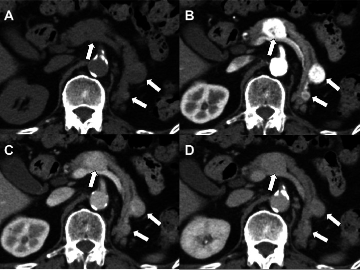
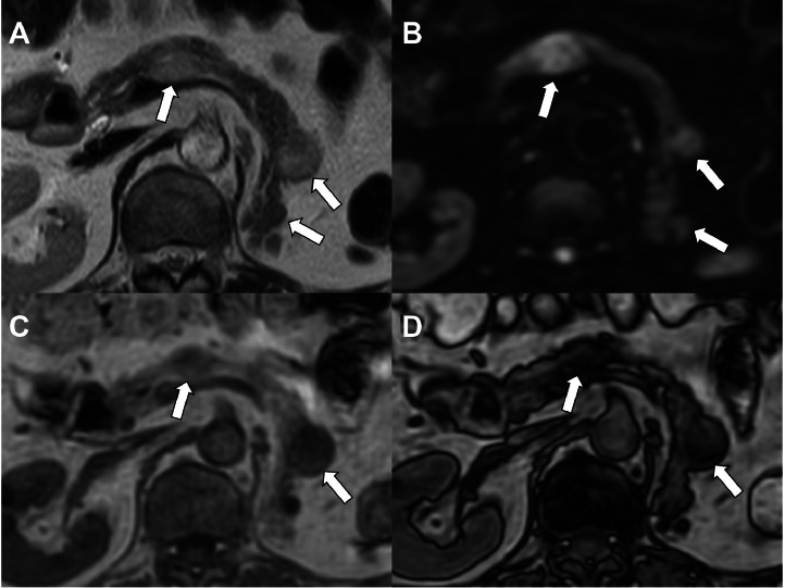
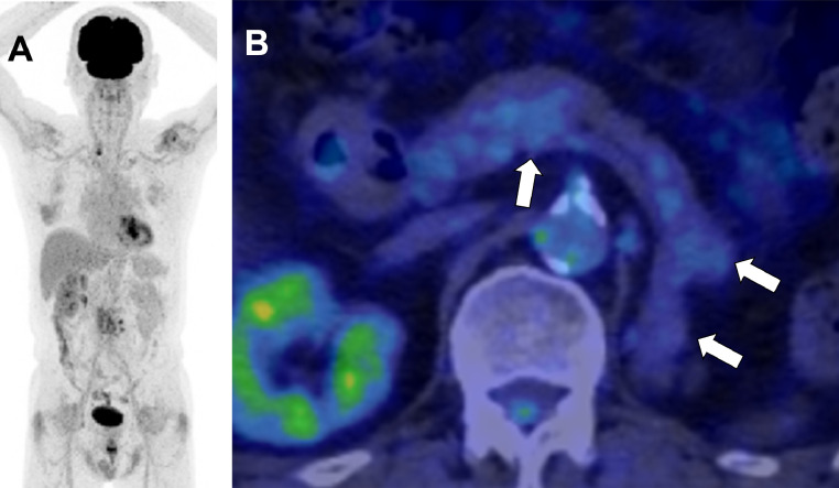
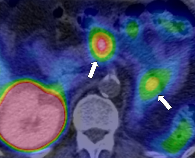
Herein, we present the case of a 73-year-old man with recurrent pancreatic tumors 20 years postnephrectomy for clear cell renal cell carcinoma (RCC). Imaging studies revealed multiple hypervascular tumors in the pancreas, posing a diagnostic challenge in differentiating RCC metastases from well-differentiated neuroendocrine tumors (NETs). Magnetic resonance imaging (MRI) chemical shift imaging played a pivotal role in suggesting the presence of fat, supporting RCC metastasis. Surgical resection confirmed the diagnosis.
Keywords: Neuroendocrine tumor; Pancreatic metastasis; Renal cell carcinoma; Somatostatin receptor scintigraphy.
© 2025 The Authors. Published by Elsevier Inc. on behalf of University of Washington.
Figures
Similar articles
-
Multiple Pancreatic Metastasis of Clear Renal Cell Carcinoma Associated with Neuroendocrine Tumor.Chirurgia (Bucur). 2023 Jun;118(3):317-323. doi: 10.21614/chirurgia.2023.v.118.i.3.p.317. Chirurgia (Bucur). 2023. PMID: 37480358
-
Repeat pancreatic resection for metachronous pancreatic metastasis from renal cell carcinoma: A case report.Int J Surg Case Rep. 2022 May;94:107022. doi: 10.1016/j.ijscr.2022.107022. Epub 2022 Apr 4. Int J Surg Case Rep. 2022. PMID: 35398785 Free PMC article.
-
Metastatic neuroendocrine pancreatic tumor - Case report.J Med Life. 2018 Jan-Mar;11(1):57-61. J Med Life. 2018. PMID: 29696066 Free PMC article.
-
Magnetic resonance imaging of pancreatic metastases from renal cell carcinoma.Clin Imaging. 2015 Nov-Dec;39(6):945-53. doi: 10.1016/j.clinimag.2015.07.012. Epub 2015 Jul 17. Clin Imaging. 2015. PMID: 26324216 Review.
-
Review of Merkel cell carcinoma with solitary pancreatic metastases mimicking primary neuroendocrine tumor of the pancreas.Clin J Gastroenterol. 2023 Oct;16(5):641-662. doi: 10.1007/s12328-023-01821-x. Epub 2023 Jul 8. Clin J Gastroenterol. 2023. PMID: 37421584 Review.
References
-
- Krenning EP, Bakker WH, Kooij PP, Breeman WA, Oei HY, de Jong M, et al. Somatostatin receptor scintigraphy with indium-111-DTPA-D-phe-1-octreotide in man: metabolism, dosimetry and comparison with iodine-123-tyr-3-octreotide. J Nucl Med. 1992;33(5):652–658. https://jnm.snmjournals.org/content/33/5/652 - PubMed
Publication types
LinkOut - more resources
Full Text Sources
