Case Reports
. 2025 Mar 8;20(5):2346-2350.
doi: 10.1016/j.radcr.2025.02.049. eCollection 2025 May.

Intracranial extension of parotid adenoid cystic carcinoma presenting as trigeminal neuralgia: A case report

Affiliations
Case Reports

Intracranial extension of parotid adenoid cystic carcinoma presenting as trigeminal neuralgia: A case report

Ammar Hassouneh et al. Radiol Case Rep. .

Abstract

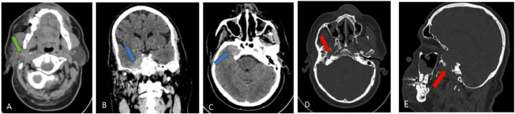
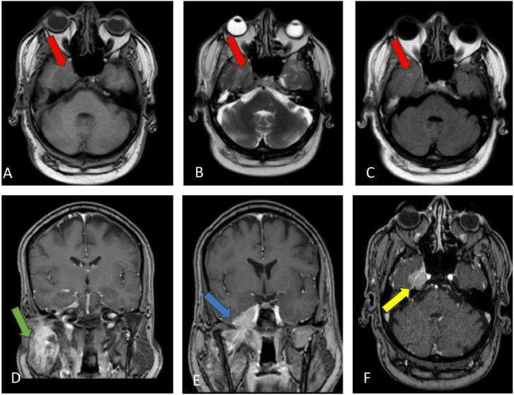
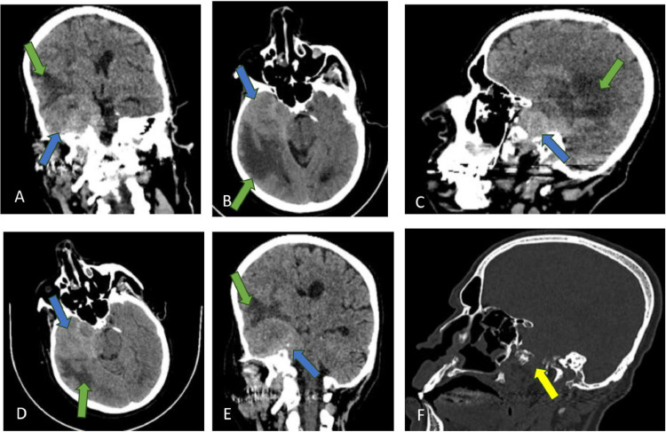
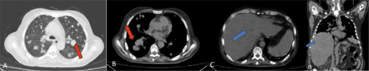
Being rare malignancies, parotid gland adenocarcinomas are most significantly represented by the ACC subtype due to their aggressive nature and propensity for PNI. We present a case of a 56-year-old male with right-sided trigeminal neuralgia and facial palsy, diagnosed with ACC of the parotid gland with intracranial extension. Tumor progression occurred with brain, lung, and liver metastases, so he was placed on palliative care despite chemotherapy. This case underscores the diagnostic and management challenges associated with ACC with PNI.

Keywords: Adenoid cystic carcinoma; Intracranial extension; Metastases; Parotid gland tumor; Perineural invasion.


Figures

Fig 1:
Fig. 1
Initial brain CT without contrast in multi planes presented in mediastinum (A) brain (B,C) and bone (D,E); (A) Enlargement of right parotid gland lesion with extension to the superficial lobe (green arrow), (B & C) Intracranial soft tissue dense lesion at the right temporal region(blue arrow), (D & E) Bone destruction at right greater wing of sphenoid (red arrow).
Fig 2:
Fig. 2
Brain MRI with and without IV contrast; (A) T1-wighted image in axial plane showed isointense lesion at the right temporal region (red arrow), (B & C) T2-wighted and FLAIR images in axial plane showed isointense lesion (red arrow), (D) T1 with IV contrast in coronal plane, showed avid enhancing lesion at the right parotid gland (green arrow), (E) T1 with IV contrast in coronal plane; showed intercranial extension of right parotid gland lesion along the mandibular nerve throw the Foramen Ovale (blue arrow), (F) T1 with IV contrast in axial plane: showed right temporal region extra axial enhancing solid mass representing intracranial extension of right parotid tumor (yellow arrow).
Fig 3:
Fig. 3
Follow up brain CT without contrast after 5 months in multi planes presented in brain (A-E) and bone (F); (A-E) Showed enlargement of previously noted intracranial extension of right parotid tumor at the right temporal region (blue arrow) with newly developed perifocal vasogenic edema (green arrow). (F) Bone destruction at right greater wing of sphenoid (yellow arrow).
Fig 4:
Fig. 4
Chest CT without IV contrast after months from first presentation; displayed in axial plane, lung (A) and mediastinum (B & C) windows and in coronal plane in mediastinum window (D); (A and B) multiple scattered lung nodules at both lung fields representing secondary deposits-metastasis (red arrow), (C and D) The visualized upper cuts for the abdomen showed enlarged liver with irregular contour occupied by multiple hypodense lesions; representing hepatic metastasis (blue arrow).

Similar articles

References

    1. Ho K., Lin H., Ann D.K., Chu P.G., Yen Y. An overview of the rare parotid gland cancer. Head Neck Oncol. 2011;3:40. doi: 10.1186/1758-3284-3-40. - DOI - PMC - PubMed
    1. de Paula F., Teshima T.H.N., Hsieh R., Souza M.M., Nico M.M.S., Lourenco S.V. Overview of Human salivary glands: highlights of morphology and developing processes. Anatom Record (Hoboken, N.J.: 2007) 2017;300(7):1180–1188. doi: 10.1002/ar.23569. - DOI - PubMed
    1. Nagao K., Matsuzaki O., Saiga H., Sugano I., Kaneko T., Katoh T., et al. Histopathologic studies on adenocarcinoma of the parotid gland. Acta Pathologica Japonica. 1986;36(3):337–347. doi: 10.1111/j.1440-1827.1986.tb01024.x. - DOI - PubMed
    1. Pinkston J.A., Cole P. Incidence rates of salivary gland tumors: results from a population-based study. Otolaryngol–Head Neck Surg. 1999;120(6):834–840. doi: 10.1016/S0194-5998(99)70323-2. - DOI - PubMed
    1. Alsanie I., Rajab S., Cottom H., Adegun O., Agarwal R., Jay A., et al. Distribution and frequency of salivary gland tumours: an international multicenter study. Head Neck Pathol. 2022;16(4):1043–1054. doi: 10.1007/s12105-022-01459-0. - DOI - PMC - PubMed

Publication types

LinkOut - more resources