Case Reports
. 2025 Mar 8;20(5):2437-2444.
doi: 10.1016/j.radcr.2025.01.089. eCollection 2025 May.

Multiple meningiomas with varying MRI features and postsurgical outcomes: A case report

Affiliations
Case Reports

Multiple meningiomas with varying MRI features and postsurgical outcomes: A case report

Rizkiditia Nugraha Hadian et al. Radiol Case Rep. .

Abstract

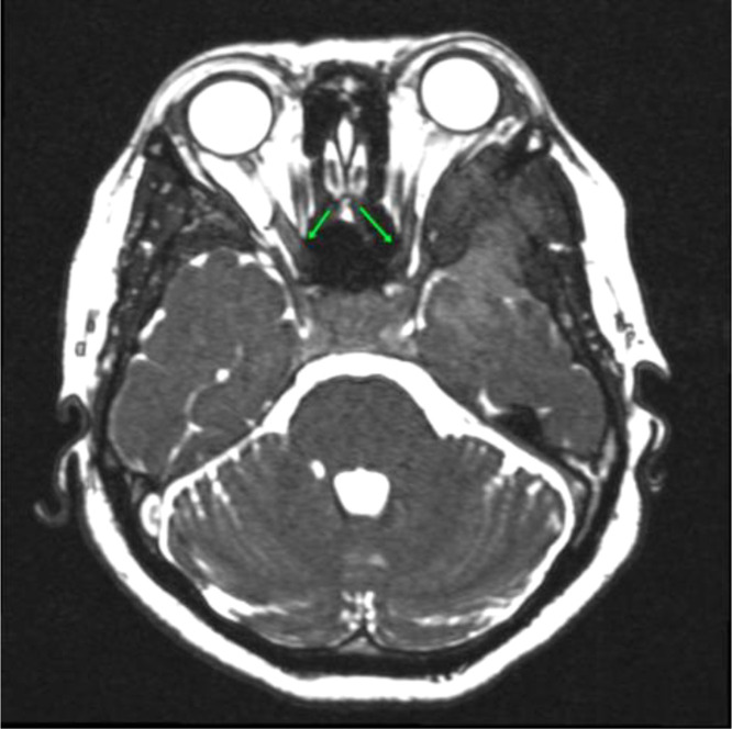
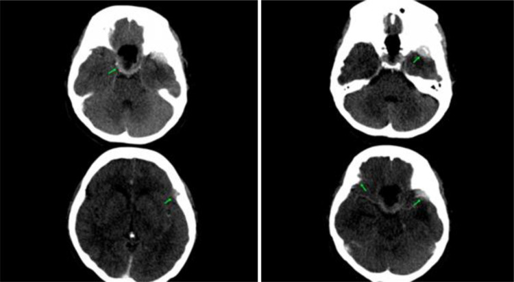
Meningiomas, which are typically benign tumors originating from the meninges, can present as multiple lesions in rare cases, occurring in 1%-10% of patients without neurofibromatosis. This report details a case involving a 49-year-old woman who initially presented with headaches followed by blurred vision, leading to the discovery of multiple meningiomas through MRI which appears as some solid mass outside the axial plane. The DWI ADC shows varying results. The patient then underwent transsphenoidal surgery for tumor resection. Histopathological analysis confirmed the presence of a meningothelial meningioma (WHO grade I) in the sellar region. Postsurgery, the patient had a CT scan showing a residual meningioma mass and experienced significant relief from her symptoms. The patient underwent outpatient treatment and was planned to undergo reoperation and follow-up MRI. The case underscores the complexity of managing multiple meningiomas. It highlights the necessity for a multidisciplinary approach to ensure accurate diagnosis and effective treatment strategies.

Keywords: Blurred vision; Brain neoplasm; Multiple meningiomas; Sphenoid tumor removal.


Figures

Fig 1
Fig. 1
The axial T1 weighted scan showed a mass in the sellar region, 2 concavities in the temporal lobes (A and B), and a concavity in the left frontal lobe (C). It also showed a CSF cleft sign (+) and a dural tail sign (+). Hyperostosis is present in both the bilateral sphenoid wing and the left frontal bone.
Fig 2
Fig. 2
Illustrates how the mass affects the optic nerve, optic chiasm, and optic tract bilaterally.
Fig 3
Fig. 3
(A) The DWI ADC examination conducted in the solid mass sellar region revealed restricted diffusion. (B) The DWI ADC examination revealed no restricted diffusion in the solid mass left temporal lobe concavity region (A) and left frontal lobe concavity region (B).
Fig 3
Fig. 3
(A) The DWI ADC examination conducted in the solid mass sellar region revealed restricted diffusion. (B) The DWI ADC examination revealed no restricted diffusion in the solid mass left temporal lobe concavity region (A) and left frontal lobe concavity region (B).
Fig 4
Fig. 4
The MRI spectroscopy examination revealed an increase in choline, glutamine, and a decrease in creatine.
Fig 5
Fig. 5
The tumor mass was removed after undergoing transsphenoidal surgery.
Fig 6
Fig. 6
The histopathologic examination revealed meningothelial meningioma (WHO grade I) at magnifications of (A) 20x, (B) 100x, and (C) 200x.
Fig 7
Fig. 7
(A) We conducted an axial brain CT scan examination using contrast after undergoing transsphenoidal surgery. A leftover mass was discovered in the sellar area, and several solid lesions outside of the axes were found in both the left frontal and temporal lobes, showing a lot of uniform enhancement. (B) A bone window head CT scan revealed defects and hyperostosis in the left frontal bone and bilateral sphenoid wing.

Similar articles

References

    1. Rogers L., Barani I., Chamberlain M., Kaley T.J., McDermott M., Raizer J., et al. Meningiomas: knowledge base, treatment outcomes, and uncertainties. A RANO review. J Neurosurg. 2015;122(1):4–23. doi: 10.3171/2014.7.JNS131644. - DOI - PMC - PubMed
    1. Bukovac A., Panić H., Mrgan T., Šlaus N., Kafka A., Njirić N., et al. Bilateral Meningioma: a case report and review of the literature. Int J Mol Sci. 2022;23(3):1187. doi: 10.3390/ijms23031187. - DOI - PMC - PubMed
    1. Lyndon D., Lansley J.A., Evanson J., Krishnan A.S. Dural masses: meningiomas and their mimics. Insights Imaging. 2019;10(1):11. doi: 10.1186/s13244-019-0697-7. - DOI - PMC - PubMed
    1. Koech F., Orege J., Ndiangui F., Macharia B., Mbaruku N. Multiple intracranial meningiomas: a review of the literature and a case report. Case Rep Surg. 2013;2013 doi: 10.1155/2013/131962. - DOI - PMC - PubMed
    1. Wong R.H., Wong A.K., Vick N., Farhat H.I. Natural history of multiple meningiomas. Surg Neurol Int. 2013;4:71. doi: 10.4103/2152-7806.112617. - DOI - PMC - PubMed

Publication types

LinkOut - more resources