

Beyond the benign: A rare case report of myxoid pleomorphic liposarcoma
- PMID: 40129807
- PMCID: PMC11930413
- DOI: 10.1016/j.radcr.2025.01.056
Beyond the benign: A rare case report of myxoid pleomorphic liposarcoma
Abstract
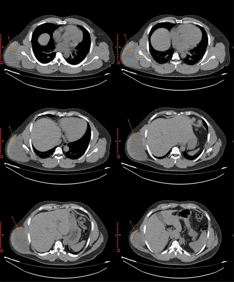
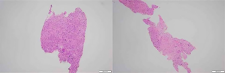
Myxoid pleomorphic liposarcoma is a rare and aggressive subtype of soft tissue sarcomas (STS). It primarily arises from adipose tissue and exhibits a high rate of recurrence and metastatic potential. We report the case of a 35-year-old male gym trainer with a 5-month history of a painless, progressively enlarging mass on the right posterior aspect of chest, diagnosed with myxoid pleomorphic liposarcoma following imaging and histopathological evaluation of the excised specimen. Surgical excision with clear margins and adjuvant radiotherapy resulted in a favorable outcome with no recurrence at 7 months. This case emphasizes the importance of early diagnosis and multidisciplinary approach in managing a rare soft tissue sarcoma to prevent complications from a delayed intervention.
Keywords: Case report; Myxoid pleomorphic liposarcoma; Oncology; Soft tissue sarcoma.
© 2025 Published by Elsevier Inc. on behalf of University of Washington.
Figures


Similar articles
-
A rare case of myxoid pleomorphic liposarcoma in an infant: A report.Int J Surg Case Rep. 2021 Oct;87:106365. doi: 10.1016/j.ijscr.2021.106365. Epub 2021 Sep 6. Int J Surg Case Rep. 2021. PMID: 34517205 Free PMC article.
-
Radiosensitivity translates into excellent local control in extremity myxoid liposarcoma: a comparison with other soft tissue sarcomas.Cancer. 2009 Jul 15;115(14):3254-61. doi: 10.1002/cncr.24375. Cancer. 2009. PMID: 19472403
-
Myxoid Pleomorphic Liposarcoma of the Spermatic Cord: A Rare Entity at a Rare Site.Cureus. 2024 Jul 28;16(7):e65554. doi: 10.7759/cureus.65554. eCollection 2024 Jul. Cureus. 2024. PMID: 39192930 Free PMC article.
-
Rare myxoid pleomorphic liposarcoma: a case report and literature review.J Clin Pathol. 2024 Apr 18;77(5):358-362. doi: 10.1136/jcp-2023-209223. J Clin Pathol. 2024. PMID: 38123348 Review.
-
Rare case of metastatic liposarcoma to the thyroid gland and a review of the literature of metastatic sarcomas to the thyroid.Head Neck. 2021 Mar;43(3):E20-E26. doi: 10.1002/hed.26552. Epub 2020 Nov 21. Head Neck. 2021. PMID: 33220159 Review.
References
-
- Siegel R.L., Miller K.D., Fuchs H.E., Jemal A. Cancer statistics, 2021. CA Cancer J Clin. 2021;71(1) - PubMed
Publication types
LinkOut - more resources
Full Text Sources
