

Distinct neural correlates of morphosyntactic and thematic comprehension processes in aphasia
- PMID: 40129862
- PMCID: PMC11930358
- DOI: 10.1093/braincomms/fcaf093
Distinct neural correlates of morphosyntactic and thematic comprehension processes in aphasia
Abstract
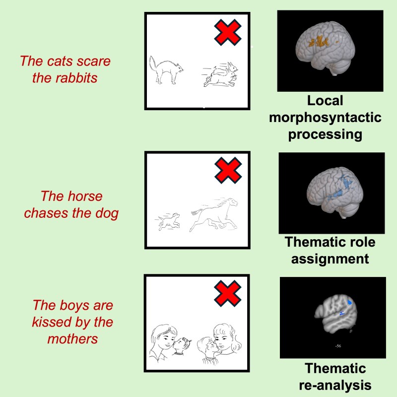
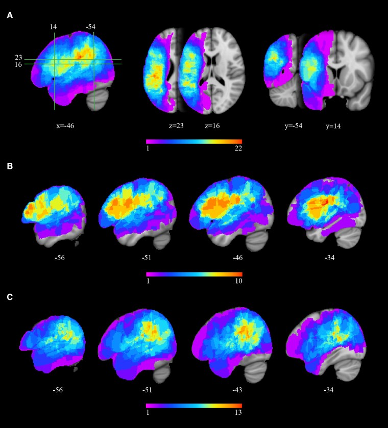
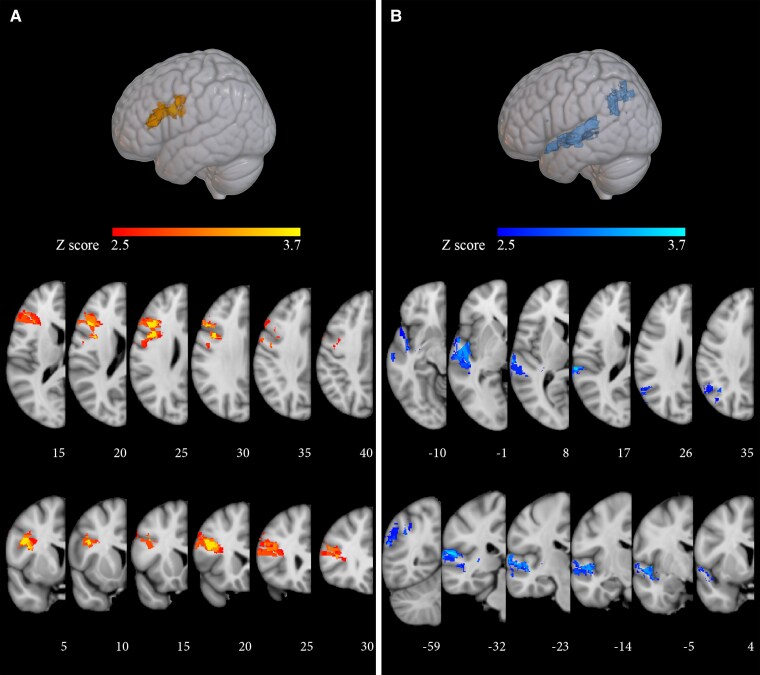
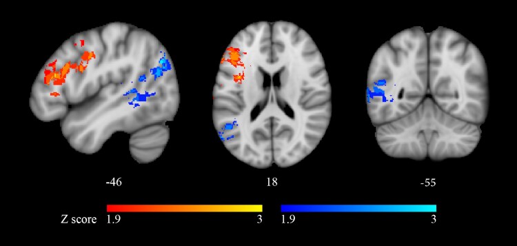
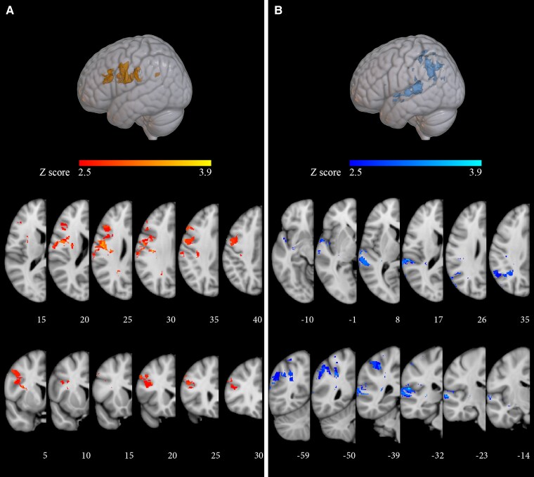
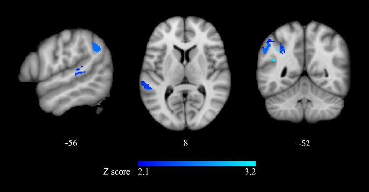
Functional neuroimaging studies in neurotypical subjects correlate sentence comprehension to a left fronto-temporo-parietal network. Recent voxel-based lesion-symptom mapping (VLSM) studies of aphasia confirm the link between sentence comprehension and a left posterior region including the angular gyrus, the supra-marginal gyrus and the postero-superior division of the temporal lobe but support left pre-frontal involvement inconsistently. However, these studies focus on thematic role assignment without considering morphosyntactic processes. Hence, available VLSM evidence could provide a partial view of the neurofunctional substrate of sentence comprehension. In the present VLSM study, both morphosyntactic and thematic processes were evaluated systematically and in the same sentence types in each participant, to provide a more detailed picture of the sentence comprehension network. Participants (33 patients with post-stroke aphasia and 90 healthy controls) completed a sentence-picture matching task in which active and passive, declarative reversible sentences were paired with morphosyntactic, thematic and lexical-semantic alternatives. Phonological short-term memory tasks were also administered. Aphasic participants were selected from an initial pool of 70 because they scored below norm on thematic foils (n = 18) or on thematic and morphological foils (n = 15), but within the norm on lexical-semantic foils. The neurofunctional correlates of morphosyntactic and thematic processes were starkly distinguishable. Pre-frontal areas including the inferior and middle frontal gyrus were involved directly in processing local morphosyntactic features and only indirectly in thematic processes. When these areas were damaged, morphosyntactic errors always co-occurred with thematic errors, probably because morphosyntactic damage disrupts the assignment of grammatical roles and ultimately that of thematic roles. Morphosyntactic errors were not influenced by word order canonicity. In contrast, selective thematic role reversals were linked to temporal and parietal damage and were significantly influenced by word order, occurring on passive more than on active sentences. An area including the angular and supra-marginal gyrus was critical for processing non-canonical word order. In sentence comprehension, pre-frontal regions are critical for processing local morphosyntactic features (at least in simple declarative sentences). Temporal and parietal regions are critical for thematic processes. Postero-superior temporal areas are involved in retrieving verb argument structure. Parietal areas are critical for assigning morphosyntactically analysed constituents to the appropriate thematic role, thus serving a crucial function in thematic re-analysis. Each area plays a prevailing but not exclusive role in these processes, interacting with other areas in the network and possibly providing both the language-specific and the domain-general resources needed at various stages of sentence comprehension.
Keywords: lesion-symptom mapping; morphosyntactic processing; sentence comprehension; thematic re-analysis; thematic role assignment.
© The Author(s) 2025. Published by Oxford University Press on behalf of the Guarantors of Brain.
Conflict of interest statement
The authors report no competing interests.
Figures
References
-
- Ferreira F. The misinterpretation of noncanonical sentences. Cogni Psychol. 2003;47(2):164–203. - PubMed
-
- Pollard C, Sag IA. Head-driven phrase structure grammar. University of Chicago Press; 1994.
-
- Bresnan J. Lexical-functional syntax. 1st ed. Wiley-Blackwell; 2000.
-
- Clifton C, Traxler MJ, Taha Mohamed M, Williams RS, Morris RK, Rayner K. The use of thematic role information in parsing: Syntactic processing autonomy revisited. J Mem and Lang. 2003;49(3):317–334.
-
- Dronkers NF, Wilkins DP, Van Valin RD, Redfern BB, Jaeger JJ. Lesion analysis of the brain areas involved in language comprehension. Cognition. 2004;92(1):145–177. - PubMed
LinkOut - more resources
Full Text Sources