. 2025 Feb 28:14:101974.
doi: 10.1016/j.toxrep.2025.101974. eCollection 2025 Jun.

Betaine alleviates deficits in motor behavior, neurotoxic effects, and neuroinflammatory responses in a rat model of demyelination

Affiliations

Betaine alleviates deficits in motor behavior, neurotoxic effects, and neuroinflammatory responses in a rat model of demyelination

Amina E Essawy et al. Toxicol Rep. .

Abstract

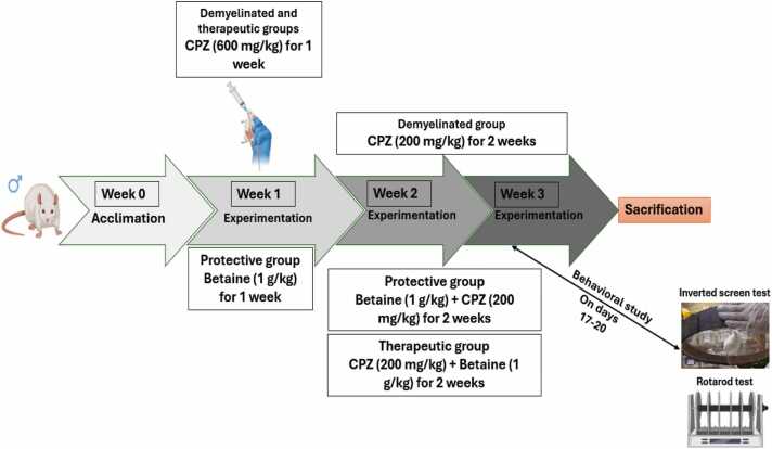
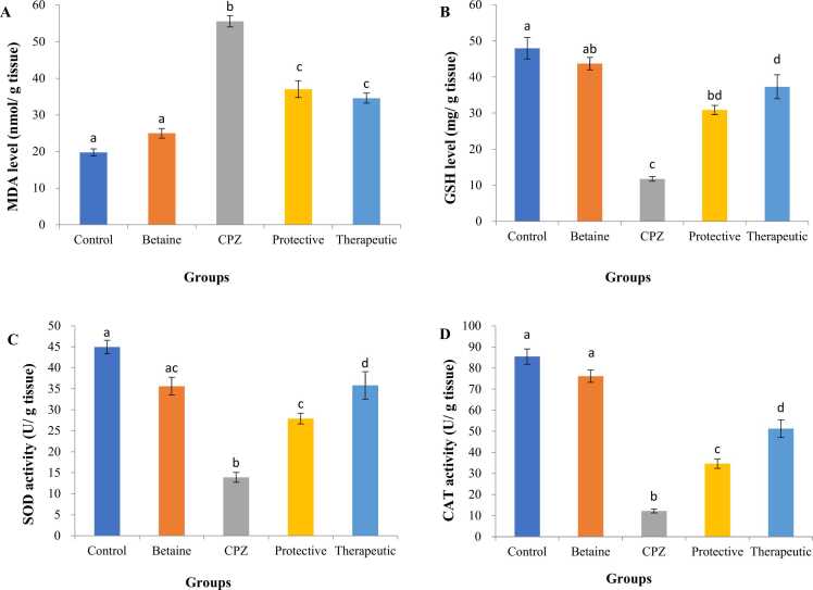
Multiple sclerosis (MS) is characterized as a chronic inflammatory demyelinating neurodegenerative disorder that leads to the deterioration of the myelin sheath and the loss of axons. Betaine, a trimethylglycine compound, is recognized for its ability to penetrate the blood-brain barrier (BBB) and exhibits properties that are antioxidant, anti-inflammatory, and neuroprotective. The cuprizone (CPZ) model serves as an effective tool for investigating the processes of demyelination and remyelination associated with MS. In our research, we examined the protective and therapeutic effects of betaine in a rat model of MS induced by CPZ. The experimental protocol involved administering 600 mg/kg of CPZ orally for 7 days, followed by 2 weeks with 200 mg/kg of CPZ. The protective group received a combination of betaine (1 g/kg/day, orally) and CPZ (200 mg/kg/day), while the therapeutic group was treated with CPZ (600 mg/kg) alongside betaine for three weeks. Behavioral assessments were conducted using inverted screen and rotarod tests to measure balance, motor coordination, and grasping ability. Following these evaluations, the rats were euthanized for analysis of oxidative stress and inflammatory biomarkers, toluidine blue staining, transmission electron microscopy (TEM) imaging, and myelin basic protein (MBP) immunostaining of the corpus callosum (CC). The results indicated that betaine significantly enhanced balance, motor coordination, and grasping ability, while decreasing oxidative stress, inhibiting interleukin (IL)-4 and IL-17 levels, and reversing the demyelination caused by CPZ. Notably, betaine also mitigated the increase in homocysteine (Hcy) levels and facilitated remyelination, evidenced by the presence of normal compacted myelin and increased expression of MBP in the CC. This study substantiates the remyelinating effects of betaine in the context of CPZ-induced demyelination. It suggests that it may contribute to the repair of myelin through the modulation of behavioral deficits, oxidative stress, neuroinflammation, ultrastructural changes, and MBP expression levels, indicating its potential as a complementary therapeutic agent in the management of MS.

Keywords: Betaine; Cuprizone; Homocysteine; Inflammation; MBP; TEM.


Conflict of interest statement

The authors declare that they have no known competing financial interests or personal relationships that could have appeared to influence the work reported in this paper.

Figures

None
Graphical abstract
Fig. 1
Fig. 1
Experimental animals and research design.
Fig. 2
Fig. 2
Effect of CPZ, betaine and their combination on the body weight changes of male rats for 3 weeks. Values are presented as mean ± S.E; n = 5 animals; mean values having different superscript letters a, b, c, d & e are significantly different from each other (P < 0.05). a, P < 0.05 vs. control. b, P < 0.05 vs. betaine. c, P < 0.05 vs. CPZ. d, P < 0.05 vs. protective (betaine + CPZ), e, P < 0.05 vs. therapeutic (CPZ+ betaine).
Fig. 3
Fig. 3
Effect of CPZ, betaine and their combination on the motor performance of male rats according to the (A) inverted screen test and (B) rotarod test. Values are presented as mean ± S.E; n = 5 animals; mean values having different superscript letters a, b, c, d & e are significantly different from each other (P < 0.05). a, P < 0.05 vs. control. b, P < 0.05 vs. betaine. c, P < 0.05 vs. CPZ. d, P < 0.05 vs. protective (betaine + CPZ), e, P < 0.05 vs. therapeutic (CPZ+ betaine).
Fig. 4
Fig. 4
Effect of CPZ, betaine and their combination on the concentration of homocysteine in the corpus callosum of male rats. Values are presented as mean ± S.E; n = 5 animals; mean values having different superscript letters a, b, c, d & e are significantly different from each other (P < 0.05). a, P < 0.05 vs. control. b, P < 0.05 vs. betaine. c, P < 0.05 vs. CPZ. d, P < 0.05 vs. protective (betaine + CPZ), e, P < 0.05 vs. therapeutic (CPZ+ betaine).
Fig. 5
Fig. 5
Effect of CPZ, betaine and their combination on the levels of (A) malondialdehyde (MDA), and (B) reduced glutathione (GSH) as well as the activities of (C) superoxide dismutase (SOD), and (D) catalase (CAT) in the corpus callosum of male rats. Values are presented as mean ± S.E; n = 5 animals; mean values having different superscript letters a, b, c, d & e are significantly different from each other (P < 0.05). a, P < 0.05 vs. control. b, P < 0.05 vs. betaine. c, P < 0.05 vs. CPZ. d, P < 0.05 vs. protective (betaine + CPZ), e, P < 0.05 vs. therapeutic (CPZ+ betaine).
Fig. 6
Fig. 6
Effect of CPZ, betaine and their combination on the levels of (A) IL-4, and (B) IL-17 in the corpus callosum of male rats. Values are presented as mean ± S.E; n = 5 animals; mean values having different superscript letters a, b, c, d & e are significantly different from each other (P < 0.05). a, P < 0.05 vs. control. b, P < 0.05 vs. betaine. c, P < 0.05 vs. CPZ. d, P < 0.05 vs. protective (betaine + CPZ), e, P < 0.05 vs. therapeutic (CPZ+ betaine).
Fig. 7
Fig. 7
Photomicrographs of semithin sections of the corpus callosum of male rats. A & B: Sections from control and betaine-treated rats respectively, showing myelinated nerve fibers with thick myelin sheath (black arrows) and glial cells with normal structure (yellow arrows). C: Section from CPZ-treated rat, showing extensively demyelinated nerve fibers (black asterisks), abnormal shaped axons (black arrows) and dense glial cells (yellow arrows). D & E: Sections from protective (betaine + CPZ) and therapeutic (CPZ+ betaine) groups showing large number of myelinated nerve fibers (black arrows), normal glial cells (yellow arrows) and few demyelinated axons (black asterisks). F & G: Light microscopical changes; % demyelination and density of the myelinated fibers, respectively in male rats' corpus callosum semithin sections stained with toluidine blue (1000X). Values are expressed as means ± S.E (n = 7); mean values having different superscript letters a, b, c, d & e are significantly different from each other (P < 0.05). a, P < 0.05 vs. control. b, P < 0.05 vs. betaine. c, P < 0.05 vs. CPZ. d, P < 0.05 vs. protective (betaine + CPZ), e, P < 0.05 vs. therapeutic (CPZ+ betaine).
Fig. 8
Fig. 8
Electron micrographs of the corpus callosum by using uranyl acetate/lead citrate stain. (A & B) Control and betaine-treated rats showing, myelinated nerve fibers with normal axons (Ax), mitochondria (M), neurofilaments and microtubules (red arrows) and myelin sheath with regular tight lamellae (thick blue arrows), an inset showing a measurement of the thickness of the myelin sheath. (C) CPZ-treated rat showing, complete demyelinated axons (DA), and other partially demyelinated axons with diffuse axonal cytoplasmic degeneration, vacuolated myelin (V), lysis in myelin sheath (dashed red arrows), splitting of myelin sheath (red arrows), axoplasm housing vacuoles (red astrikes), and mitochondria with indistinct cristae (M). Axons almost all of them have irregular deformed myelin sheaths, an inset showing a measurement of the thickness of the myelin sheath. (D & E) betaine + CPZ (protective group) and CPZ + betaine (therapeutic group) showing, intact axons (Ax) with marked myelination, neurofilaments and microtubules (red arrows), mitochondria (M), and myelin sheath with regular tight lamellae (thick blue arrows). Few demyelinated nerve fibers (DA) are also encountered, an inset showing a measurement of the thickness of the myelin sheath. Microscopic magnification: 2 µm, inset 200 nm. (F) Axon diameter, (G) fiber diameter, (H) myelin thickness, and (I) G ratio as demonstrated by TEM imaging of the corpus callosum. Data presented as mean ± S.E (n = 10); mean values having different superscript letters a, b, c, d & e are significantly different from each other (P < 0.05). a, P < 0.05 vs. control. b, P < 0.05 vs. betaine. c, P < 0.05 vs. CPZ. d, P < 0.05 vs. protective (betaine + CPZ), e, P < 0.05 vs. therapeutic (CPZ+ betaine).
Fig. 8
Fig. 8
Electron micrographs of the corpus callosum by using uranyl acetate/lead citrate stain. (A & B) Control and betaine-treated rats showing, myelinated nerve fibers with normal axons (Ax), mitochondria (M), neurofilaments and microtubules (red arrows) and myelin sheath with regular tight lamellae (thick blue arrows), an inset showing a measurement of the thickness of the myelin sheath. (C) CPZ-treated rat showing, complete demyelinated axons (DA), and other partially demyelinated axons with diffuse axonal cytoplasmic degeneration, vacuolated myelin (V), lysis in myelin sheath (dashed red arrows), splitting of myelin sheath (red arrows), axoplasm housing vacuoles (red astrikes), and mitochondria with indistinct cristae (M). Axons almost all of them have irregular deformed myelin sheaths, an inset showing a measurement of the thickness of the myelin sheath. (D & E) betaine + CPZ (protective group) and CPZ + betaine (therapeutic group) showing, intact axons (Ax) with marked myelination, neurofilaments and microtubules (red arrows), mitochondria (M), and myelin sheath with regular tight lamellae (thick blue arrows). Few demyelinated nerve fibers (DA) are also encountered, an inset showing a measurement of the thickness of the myelin sheath. Microscopic magnification: 2 µm, inset 200 nm. (F) Axon diameter, (G) fiber diameter, (H) myelin thickness, and (I) G ratio as demonstrated by TEM imaging of the corpus callosum. Data presented as mean ± S.E (n = 10); mean values having different superscript letters a, b, c, d & e are significantly different from each other (P < 0.05). a, P < 0.05 vs. control. b, P < 0.05 vs. betaine. c, P < 0.05 vs. CPZ. d, P < 0.05 vs. protective (betaine + CPZ), e, P < 0.05 vs. therapeutic (CPZ+ betaine).
Fig. 9
Fig. 9
Electron micrographs of the corpus callosum by using uranyl acetate/lead citrate stain. (A & B) Control and betaine-treated rats showing, an oligodendrocyte having a large nucleus (N) with clumped peripheral heterochromatin and bilayer nuclear envelope (white arrow), and dense cytoplasm containing mitochondria (M), Golgi apparatus (G), short cisternae of rough endoplasmic reticulum (rER) and some dense inclusions. (C) CPZ-treated rat showing an oligodendrocyte with pyknotic hyperchromatic nucleus (N), dilated nuclear envelope (white arrow) and degenerated mitochondria (M). (D & E) betaine + CPZ (protective group) and CPZ + betaine (therapeutic group) showing an oligodendrocyte having a large, rounded nucleus (N) with well peripheral heterochromatin and distinct nuclear envelope (white arrow), and dense cytoplasm containing mitochondria (M), Golgi apparatus (G) and short cisternae of rough endoplasmic reticulum (rER) and some dense inclusions. Scale bar (1 µm), inset 200 nm.
Fig. 10
Fig. 10
Immunohistochemistry showing the expression of MBP in the CC of rats in the five experimental groups: (A) Control, (B) Betaine, (C) CPZ, (D) Betaine + CPZ, (E) CPZ + Betaine. MPB positive cells (brown staining) decreases dramatically in CPZ group. Both protective (betaine + CPZ) and therapeutic (CPZ + betaine)-treated groups can reverse the reduction in MBP expression in the CC. Scale bar: 50 µm.
Fig. 11
Fig. 11
(A) Spearman correlation analysis, representing behavioral, weight, Hcy concentration, oxidant/antioxidant, and inflammatory biomarkers, as well as semithin section and ultrastructural changes, are superimposed as supplementary variables. (B) The analysis of protein-protein interactions involving variously expressed redox and inflammatory markers in relation to myelin basic protein utilizing the STRING database.

Similar articles

References

    1. Abd El Aziz A.E., Sayed R.H., Sallam N.A., El Sayed N.S. Neuroprotective effects of telmisartan and nifedipine against cuprizone-induced demyelination and behavioral dysfunction in mice: roles of NF-κB and Nrf2. Inflam. 2021;44:1629–1642. doi: 10.1007/s10753-021-01447-6. - DOI - PubMed
    1. Abdel-Maged A.E., Gad A.M., Rashed L.A., Azab S.S., Mohamed E.A., Awad A.S. Repurposing of secukinumab as neuroprotective in cuprizone-induced multiple sclerosis experimental model via inhibition of oxidative, inflammatory, and neurodegenerative signaling. Mol. Neurobiol. 2020;57:3291–3306. doi: 10.1007/s12035-020-01972-9. - DOI - PubMed
    1. Acs P., Komoly S. Selective ultrastructural vulnerability in the cuprizone-induced experimental demyelination. Ideggyogy. Sz. 2012;65:266–270. - PubMed
    1. Aharoni R., Eilam R., Arnon R. Astrocytes in multiple sclerosis—Essential constituents with diverse multifaceted functions. Int. J. Mol. Sci. 2021;22 doi: 10.3390/ijms22115904. - DOI - PMC - PubMed
    1. Aldhahri R.S., Alghamdi B.S., Alzahrani N.A., Bahaidrah K.A., Alsufiani H.M., Mansouri R.A., Ashraf G.M. Biochanin A improves memory decline and brain pathology in cuprizone-induced mouse model of multiple sclerosis. Behav. Sci. 2022;12:1–18. doi: 10.3390/bs12030070. - DOI - PMC - PubMed

LinkOut - more resources