. 2025 Feb 27:9:100267.
doi: 10.1016/j.bioflm.2025.100267. eCollection 2025 Jun.

From adhesion to biofilms formation and resilience: Exploring the impact of silver nanoparticles-based biomaterials on Pseudomonas aeruginosa

Affiliations

From adhesion to biofilms formation and resilience: Exploring the impact of silver nanoparticles-based biomaterials on Pseudomonas aeruginosa

Maya Rima et al. Biofilm. .

Abstract

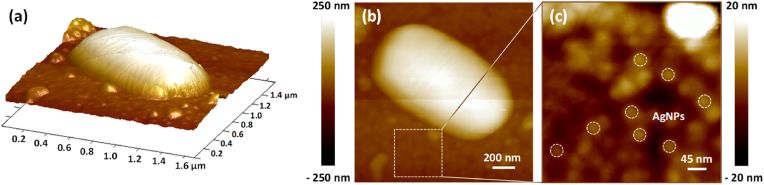
Colonization of medical devices by microorganisms, often progressing to the formation of resilient biofilms, presents a common clinical issue. To address this challenge, there is growing interest in developing novel biomaterials with antimicrobial/antibiofilm properties as a promising preventive measure. This study explores nanocomposite biomaterials based on silver nanoparticles (AgNPs) deposited on thin silica (SiO2) layers for their potential effect on the adhesion, detachment, viability and biofilm formation of the opportunistic Pseudomonas aeruginosa. The AgNPs-based biointerface effect on biofilm development is investigated on the PAO1-Tn7-gfp strain by combining experiments under static and dynamic conditions. For the latter, a shear-stress flow chamber is used to mimic conditions encountered around certain medical devices. The findings reveal a rapid bactericidal effect of the AgNPs, noticeable within 30 min of exposure. Moreover, a delay in surface colonization is observed with a thin and unstructured biofilm, even after 72h of dynamic culture. A considerable fragility and sensitivity to hydrodynamic stresses is noticed for this loosely attached bacterial monolayer when compared with the thick and resilient biofilm formed on SiO2 surface. This study underlines the potential of AgNPs-based biomaterials in the conception of novel antimicrobial/antibiofilm surfaces with controlled release of the biocidal agent.

Keywords: Antiadhesion; Antibiofilm; Antimicrobial; Biointerfaces; Pseudomonas aeruginosa; Silver nanoparticle-based biomaterials; plasma (gas discharge) process.


Conflict of interest statement

The authors declare the following financial interests/personal relationships which may be considered as potential competing interests: Kremena Makasheva reports financial support was provided by 10.13039/501100001665French National Research Agency. If there are other authors, they declare that they have no known competing financial interests or personal relationships that could have appeared to influence the work reported in this paper.

Figures

Fig. 1
Fig. 1
Schematic representation of the implemented strategy to evaluate the surface colonization phases by P. aeruginosa PAO1-Tn7-gfp, starting from adhesion to biofilm formation and resilience, when in contact with AgNPs: AgNPs-based biomaterials, composed of a single layer of AgNPs deposited on thin SiO2 layer thermally grown on Si-substrate (a), were assessed across the different stages of bacterial biofilm formation, under static and dynamic conditions. This holistic approach encompasses adhesion (b), adhesion strength (c), biofilm formation (d), and biofilm resilience (e).
Fig. 2
Fig. 2
Schematic representation of the shear-stress flow chamber and the experimental set-up employed in the biofilm experiments.
Fig. 3
Fig. 3
Structural characterization of the AgNPs-based biomaterials. (a, b) SEM top-view images at two resolution scales, (c) histogram showing results from statistics performed on 222 AgNPs: size distribution by bins and the corresponding Gaussian distribution function, (d) AFM topography image of the sample surface, including the arithmetic and quadratic surface height parameters, (e) AFM surface topography profile along 6 AgNPs, as marked with the white line on the topography image, and (f) extracted high order surface height parameters (skewness and kurtosis) of the studied area.
Fig. 4
Fig. 4
Study of the adhesion and viability of P. aeruginosa PAO1-Tn7-gfp on SiO2-samples and AgNPs-based biomaterials. Cultivable planktonic and adhered bacteria were quantified after 30 and 90 min of contact under static conditions. Results are expressed as means log CFU/cm2 ± Standard Deviation (SD) from three independent experiments. In accordance with the applied methodology for counting the planktonic cells log CFU/cm2 = log CFU/mL. Statistically significant differences are determined by two-way ANOVA with Tukey’ test for multiple comparisons (∗∗, p-value <0.01, ∗∗∗∗, p-value <0.0001), ns: not significant, nd: not detected. Epifluorescence microscopic images are presented in Supplementary Materials, Fig. S1.
Fig. 5
Fig. 5
Percentage of live and damaged/dead P. aeruginosa PAO1-Tn7-gfp cells on SiO2-samples and AgNPs-based biomaterials at t0 (just after bacterial injection) and after 30 and 90 min of contact under static conditions in WIP in the shear-stress flow chamber (a). Wall shear-stress induced detachment profiles of P. aeruginosa PAO1-Tn7-gfp adhered on SiO2-samples and AgNPs-based biomaterials (b), (the red dash line on the figure shows the 50 % limit). Results are expressed as means ± SD from three independent experiments. Statistically significant differences determined by two-way ANOVA with Tukey's test for multiple comparisons (∗, p-value < 0.05) between t90 min and after wall shear-stress application are indicated. (For interpretation of the references to color in this figure legend, the reader is referred to the Web version of this article.)
Fig. 6
Fig. 6
Biofilm formation by PAO1-Tn7-gfp with the corresponding fluorescence intensity (FI) (magnification x20, resolution 2.7 pixels/micron) on SiO2-samples and AgNPs-based biomaterials after 24 (a, b, c), 48 (d, e, f), and 72h (g, h, i) of dynamic culture. FI results for both live and damaged/dead bacteria are expressed as means ± SD from three independent experiments. Statistically significant differences determined by two-way ANOVA with Tukey’ test for multiple comparisons (∗∗∗, p-value <0.001) between SiO2-samples and AgNPs-based biomaterials are indicated. nd: not detected.
Fig. 7
Fig. 7
AFM topography images, in tapping mode, of 72h-old biofilm formed on SiO2-samples in 3D (6 μm × 6 μm) in (a) and 2D (32 μm × 32 μm) in (b) representations. The respective heights and occurrences are shown in (c) and (d). The displayed height profile in (c) was determined along the white dashed line shown in (b) The occurrence diagram in (d) was determined over the entire topography map in (b).
Fig. 8
Fig. 8
AFM topography images, in tapping mode, of AgNPs-based biomaterials after 72h of dynamic culture in 3D (1.6 μm × 1.6 μm) on (a) and 2D (1.6 μm × 1.6 μm) on (b) representations; the height color bar is common for the two representations, in (a) and (b). Magnification (425 nm × 425 nm) of the dashed square on (b), representing the remaining AgNPs (some of the AgNPs are suggested by the white dashed circles) is given on (c). (For interpretation of the references to color in this figure legend, the reader is referred to the Web version of this article.)
Fig. 9
Fig. 9
Epifluorescence microscopy images of P. aeruginosa PAO1-Tn7- gfp 72h-old biofilms formed on SiO2-samples (a), after application of low (0.3 Pa), medium (4.3 Pa) and very high (16.7 Pa) wall shear-stresses. Shear-flow induced detachment profiles of live and dead bacteria on SiO2-samples (b). The same sequence but for AgNPs-based biomaterials: epifluorescence microscopy images of P. aeruginosa PAO1-Tn7- gfp 72h-old biofilms (c) and wall shear-stress induced detachment profiles of live and dead bacteria (d).

References

    1. Mehta A.C., Muscarella L.F. Bronchoscope-related "superbug" infections. Chest. 2020;157(2):454–469. doi: 10.1016/j.chest.2019.08.003. - DOI - PubMed
    1. Zhang Y., Zhou H., Jiang Q., Wang Q., Li S., Huang Y. Bronchoscope-related Pseudomonas aeruginosa pseudo-outbreak attributed to contaminated rinse water. Am J Infect Control. 2020;48(1):26–32. doi: 10.1016/j.ajic.2019.06.013. - DOI - PubMed
    1. Kuehl R., Brunetto P.S., Woischnig A.K., Varisco M., Rajacic Z., Vosbeck J., Terracciano L., Fromm K.M., Khanna N. Preventing implant-associated infections by silver coating. Antimicrob Agents Chemother. 2016;60(4):2467–2475. doi: 10.1128/AAC.02934-15. - DOI - PMC - PubMed
    1. Arciola C.R., Campoccia D., Montanaro L. Implant infections: adhesion, biofilm formation and immune evasion. Nat Rev Microbiol. 2018;16(7):397–409. doi: 10.1038/s41579-018-0019-y. - DOI - PubMed
    1. Ul Haq I., Khan T.A., Krukiewicz K. Etiology, pathology, and host-impaired immunity in medical implant-associated infections. J Infect Public Health. 2024;17(2):189–203. doi: 10.1016/j.jiph.2023.11.024. - DOI - PubMed

LinkOut - more resources