. 2025 Mar 10:49:207-217.
doi: 10.1016/j.bioactmat.2025.03.001. eCollection 2025 Jul.

Engineering a stem cell-embedded bilayer hydrogel with biomimetic collagen mineralization for tendon-bone interface healing

Affiliations

Engineering a stem cell-embedded bilayer hydrogel with biomimetic collagen mineralization for tendon-bone interface healing

Tingyun Lei et al. Bioact Mater. .

Abstract

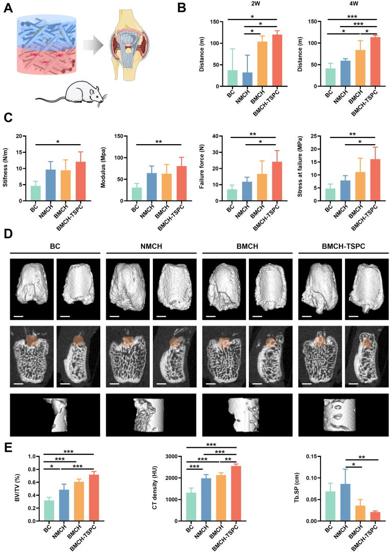
The tendon-bone interface effectively transfers mechanical stress for movement, yet its regeneration presents significant clinical challenges due to its hierarchical structure and composition. Biomimetic strategies that replicate the distinctive characteristics have demonstrated potential for enhancing the healing process. However, there remains a challenge in developing a composite that replicates the nanostructure of the tendon-bone interface and embeds living cells. Here, we engineered a nanoscale biomimetic bilayer hydrogel embedded with tendon stem cells for tendon-bone interface healing. Specifically, the biomimetic hydrogel incorporates intra- and extrafibrillar mineralized collagen fibrils as well as non-mineralized collagen fibrils resembling the tendon-bone interface at the nanoscale. Furthermore, biomimetic mineralization with the presence of cells realizes living tendon-bone-like tissue constructs. In the in vivo patella-patellar tendon-interface injury model, the tendon stem cell-laden biomimetic hydrogel promoted tendon-bone interface regeneration, demonstrated by increased fibrocartilage formation, improved motor function, and enhanced biomechanical outcomes. This study highlights the potential of the stem cell-laden biomimetic hydrogel as an effective strategy for tendon-bone interface regeneration, offering a novel approach to engineering complex tissue interfaces.

Keywords: Biomimetic mineralization; Hydrogel; Tendon stem cell; Tendon-bone interface.


Conflict of interest statement

The authors declare that they have no known competing financial interests or personal relationships that could have appeared to influence the work reported in this paper.

Figures

Image 1
Graphical abstract
Fig. 1
Fig. 1
Fabrication and characterization of the biomimetic hydrogel. (A) Schematic illustration of the biomimetic hydrogel fabrication process. (B) Optical and Alizarin red staining images of the bilayer collagen hydrogel. Scale bar = 1 mm. (C) Thermogravimetric analysis of the mineralized collagen hydrogel and non-mineralized collagen hydrogel. (D) Elastic modulus of the mineralized and non-mineralized collagen hydrogel. (E) FTIR spectra of the mineralized and non-mineralized collagen hydrogels. The green dashed line represented the peak position of amide I, II, and III. The orange dashed line represented the peak position of apatitic phosphate. (F) TEM images and SAED of mineralized and non-mineralized collagen. Scale bar = 1 μm (left), 200 nm (right). (G) Cryo-SEM images of mineralized and non-mineralized collagen. Scale bar = 5 μm (left), 3 μm (right). (H) EDX spectra of mineralized and non-mineralized collagen. (I) STORM image of the mineralized collagen fibrils. Scale bar = 100 nm. Non-min, non-mineralized. Min, mineralized.
Fig. 2
Fig. 2
Analysis of mineralization-induced biological changes. (A) Schematic diagram of cell-laden non-mineralized and mineralized hydrogel. (B) Representative 3D live/dead fluorescence images. Scale bar = 100 μm. (C) Representative fluorescence and SEM images of cell morphology. Aspect and area of F-actin stained cells. Scale bar = 50 μm (fluorescence images), 30 μm (SEM images). (D) PCA plot showing distinct expression patterns. (E) Heatmap showing pearson correlation. (F) Heatmap showing differentially expressed genes between mineralized and non-mineralized group. (G) Heatmap showing the level of gene expression. (H) GO analysis of upregulated genes in mineralized group. (I) KEGG analysis of upregulated genes in mineralized group. Non-min, non-mineralized. Min, mineralized.
Fig. 3
Fig. 3
Cell-laden biomimetic hydrogel promoted functional recovery and new bone formation. (A) Schematic depicts the cell-laden biomimetic hydrogel for tendon-bone interface healing. (B) Running distances of rats at 2 and 4 weeks post-surgery. (C) Biomechanical tests of the repaired interface tissues from different groups at 4 weeks post-surgery. (D) Representative micro-CT images of different groups. Sagittal and coronal-sections, along with 3D reconstruction of micro-CT images, from the specified region of different groups. Scale bar = 1 mm. (E) Quantitative analysis of bone formation in the specified region.
Fig. 4
Fig. 4
Cell-laden biomimetic hydrogel promoted histological structure restoration. (A) Representative histological staining images (H&E, SO-FG, TB and Masson) of different groups. Scale bar = 100 μm. (B) Representative immunofluorescence staining images for COL2 and SOX9 of different groups. Scale bar = 50 μm. (C) Histological score, the area of newly formed fibrocartilage, and the area of COL2 of different groups.
Fig. 5
Fig. 5
scRNA-seq analysis of the repaired interface tissues in BMCH-TSPC group. (A) Uniform manifold approximation and projection (UMAP) plot showing the unbiased clustering results. (B) UMAP plot showing the unbiased clustering of tendon-bone cell lineages. (C) Predicted differentiation stages for each cluster. (D) Heatmap showing DEG in each major cluster (left panel). Bubble diagram showing enriched GO terms (right panel). (E) Dotplot showing gene expression levels across clusters. (F) Differentiation trajectory of subpopulations predicted by Monocle2. (G) Heatmap showing gene expressions profiles along the pseudotime of two distinct trajectories (left panel). Expression of genes visualized on differentiation trajectory (right panel). (H) Representative H&E and SO-FG staining images at 2 weeks post-surgery. Scale bar = 100 μm. (I) Representative immunofluorescence staining images for COL2 and SOX9 at 2 weeks post-surgery. Scale bar = 100 μm.

References

    1. Golman M., Abraham A.C., Kurtaliaj I., Marshall B.P., Hu Y.J., Schwartz A.G., Guo X.E., Birman V., Thurner P.J., Genin G.M., Thomopoulos S. Toughening mechanisms for the attachment of architectured materials: the mechanics of the tendon enthesis. Sci. Adv. 2021;7 doi: 10.1126/sciadv.abi5584. eabi5584. - DOI - PMC - PubMed
    1. Font Tellado S., Balmayor E.R., Van Griensven M. Strategies to engineer tendon/ligament-to-bone interface: biomaterials, cells and growth factors. Adv. Drug Deliv. Rev. 2015;94:126–140. doi: 10.1016/j.addr.2015.03.004. - DOI - PubMed
    1. Bedi A., Bishop J., Keener J., Lansdown D.A., Levy O., MacDonald P., Maffulli N., Oh J.H., Sabesan V.J., Sanchez-Sotelo J., Williams R.J., Feeley B.T. Rotator cuff tears. Nat. Rev. Dis. Primers. 2024;10 doi: 10.1038/s41572-024-00492-3. - DOI - PubMed
    1. Shengnan Q., Bennett S., Wen W., Aiguo L., Jiake X. The role of tendon derived stem/progenitor cells and extracellular matrix components in the bone tendon junction repair. Bone. 2021;153 doi: 10.1016/j.bone.2021.116172. - DOI - PubMed
    1. Zhao S., Su W., Shah V., Hobson D., Yildirimer L., Yeung K.W.K., Zhao J., Cui W., Zhao X. Biomaterials based strategies for rotator cuff repair. Colloids Surf. B Biointerfaces. 2017;157:407–416. doi: 10.1016/j.colsurfb.2017.06.004. - DOI - PubMed

LinkOut - more resources