. 2025 Feb;10(2):187-199.
doi: 10.1016/j.jacbts.2024.09.009. Epub 2024 Nov 27.

Endothelial JCAD Worsens Acute Ischemic Stroke Outcomes by Enhancing Inflammation in Response to Ischemia/Reperfusion

Affiliations

Endothelial JCAD Worsens Acute Ischemic Stroke Outcomes by Enhancing Inflammation in Response to Ischemia/Reperfusion

Stefano Ministrini et al. JACC Basic Transl Sci. 2025 Feb.

Abstract

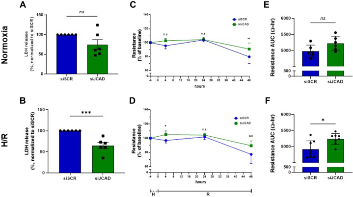
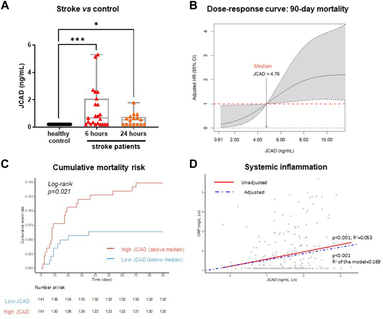
The role of junctional protein associated with coronary artery disease (JCAD) in acute ischemic stroke (AIS) has not been investigated yet. To investigate its potential as a therapeutic target, transient middle cerebral artery occlusion was induced in JCAD knockout mice, with improvement of stroke outcome and reduced blood-brain barrier permeability and expression of vascular cell adhesion molecule (VCAM)-1. JCAD plays a deleterious role in ischemia/reperfusion cerebral damage and associates with higher 90-day mortality in patients with AIS. JCAD may thus represent a novel prognostic biomarker for patients with AIS, as well as a therapeutic target.

Keywords: acute ischemic stroke; blood-brain barrier; endothelial cells; hypoxia/reoxygenation; inflammation.


Conflict of interest statement

Funding Support and Author Disclosures This work was funded by Swiss Heart Foundation grant FF22014/2022 (Dr Ministrini), the Novartis Foundation for Medical-Biological Research grant 21B070 (Prof Liberale), and Swiss National Science Foundation grant 501100001711-197510] (Prof Camici). Dr Puspitasari is the recipient of a Forschungskredit Candoc grant from the University of Zurich and a grant from Swiss Life Foundation for Public Health and Medical Research. Dr Kraler has received institutional research grants from the Jubiläumsstiftung SwissLife, the Lindenhof Foundation, the Novartis Foundation for Medical-Biological Research, the Swiss Heart Foundation, the Swiss Society of Cardiology, and the Theodor-Ida-Herzog-Egli Foundation; has received equipment and materials from Roche Diagnostics outside the submitted work; and has received travel support from the European Atherosclerosis Society, the European Society of Cardiology, the European Society of Clinical Investigation, Sphingotec GmbH, the 4TEEN4 Pharmaceuticals GmbH, and PAM Theragnostics GmbH. Dr Wenzl has received financial support from the Foundation for Cardiovascular Research-Zurich Heart House, the Lindenhof Foundation, the European Society of Cardiology, the Swiss Heart Foundation, the Fonds zur Förderung des akademischen Nachwuchses of the University of Zurich, the Medical University of Graz, the Theodor-Ida-Herzog-Egli Foundation, the Sphingotec GmbH, the 4TEEN4 Pharmaceuticals GmbH, and the PAM Theragnostics GmbH outside this work. Prof Katan has received funding from the Swiss National Science Foundation (Project Nr. 182267 and Project Nr. 213471), the Swiss Heart Foundation, and in kind contributions from ROCHE Diagnostics and BRHAMS Thermofisher, outside the submitted work. Prof Montecucco is the recipient of Rete CARDIOLOGICA grant RCR-2022-23682288. Work supported by #NEXTGENERATIONEU (NGEU) and funded by the Ministry of University and Research (MUR), National Recovery and Resilience Plan (NRRP), project MNESYS (PE0000006) - (DN. 1553 11.10.2022) to Prof. Montecucco Prof Lüscher holds leadership positions at the European Society of Cardiology, Swiss Heart Foundation, and the Foundation for Cardiovascular Research-Zurich Heart House; has received institutional educational and research grants outside this work from Abbott, Amgen, AstraZeneca, Boehringer Ingelheim, Daichi Sankyo, Eli Lilly, Novartis. Novo Nordisk, Sanofi, Servier, and Vifor; and has received consulting fees from Dacadoo, Novartis, Novo Nordisk, Pfizer, and Philips. Prof Camici and Prof Liberale are coinventors on the International Patent WO/2020/226993 filed in April 2020; the patent relates to the use of antibodies which specifically bind interleukin-1a to reduce various sequelae of ischemia-reperfusion injury to the central nervous system. Prof Camici is a consultant to Sovida Solutions limited; is the recipient of a Sheikh Khalifa’s Foundation Assistant Professorship at the Faculty of Medicine, University of Zurich; and has received financial support by the Alfred and Annemarie von Sick Grants for Translational and Clinical Research Cardiology and Oncology and by the Swiss Heart Foundation. Prof Liberale has received financial support from the Swiss Heart Foundation and the Novartis Foundation for Medical-Biological Research. All other authors have reported that they have no relationships relevant to the contents of this paper to disclose.

Figures

None
Graphical abstract
Figure 1
Figure 1
Role of JCAD Expression in a Murine Model of Acute Brain Ischemia/Reperfusion by Transient Middle Cerebral Artery Occlusion Effect of total JCAD knockout (ko) (Jcad−/−) on ischemic volume (A and B), motor coordination, assessed by RotaRod test (C), and neurological status, assessed by Bederson scale score (D) Wild-type (Jcad+/+) littermates were used as control mice; n = 7 Jcad−/− mice; n = 6 Jcad+/+ mice. Effect of endothelial-specific JCAD knockout (eJcad−/−) on ischemic volume (E and F), motor coordination (G), and neurological status (H). Jcad “floxed” (Jcadfl/fl) littermates were used as control mice; n = 9 eJcad−/− mice; n = 7 Jcadfl/fl mice. Effect of postischemic JCAD silencing by small interfering RNA (siJCAD) on ischemic volume (I and J), motor coordination (K), and neurological status (L). RNA scramble (siSCR)-treated littermates were used as control mice; n = 7 siJCAD mice; n = 7 siSCR mice. Data are presented as mean ± SEM. Student’s t-test (A, E, I) and 2-way repeated measures analysis of variance with Sidak’s correction for multiple comparisons (C, D, G,H, K, and L); ∗P < 0.05; ∗∗P < 0.01; and ∗∗∗P < 0.001.
Figure 2
Figure 2
Effect of H/R Injury in Human Brain Microvascular Endothelial Cells After JCAD Silencing Cells were exposed to normoxia (A, C, and E) or hypoxia/reoxygenation (H/R) injury (B, D, and F). Cell death rate, assessed as lactic dehydrogenase (LDH) release in silenced (siJCAD) and unsilenced (siSCR) cells (A and B). Endothelial monolayer integrity, assessed as trans-endothelial electric resistance, in silenced and unsilenced cells (C and D, and E and F). Data are presented as mean ± SEM. n = 6, Student’s t-test (A, B, E, and F) and 2-way repeated measures analysis of variance with Sidak’s correction for multiple comparisons (C and D); ∗P < 0.05; ∗∗P < 0.01; and ∗∗∗P < 0.001. AUC = area under the curve; other abbreviations as in Figure 1.
Figure 3
Figure 3
Molecular Effects of JCAD Silencing in Human Brain Microvascular Endothelial Cells Exposed to Nx or H/R Injury Protein expression was assessed by Western Blot. Vascular endothelial cadherin (VE-cadherin) (A), occludin (B), claudin 5 (C), vascular cell adhesion molecule (VCAM)-1 (D), and intercellular adhesion molecule (ICAM)-1 (E). Data are presented as mean ± SEM. n = 6, 1-way analysis of variance with Tukey’s correction for multiple comparisons; ∗P < 0.05; ∗∗P < 0.01; and ∗∗∗P < 0.001. Nx = normoxia; other abbreviations as in Figures 1 and 2.
Figure 4
Figure 4
Role of Endothelial JCAD Expression in the Pathophysiology of Acute Brain Ischemia Acute brain ischemia has been reproduced in mice with endothelial-specific JCAD deletion (eJcad−/−) by transient middle cerebral artery occlusion. Effect of endothelial JCAD deletion on blood-brain barrier integrity, assessed by extravascular IgG immunostaining (A and B) after transient middle cerebral artery occlusion. Effect of endothelial JCAD deletion on endothelial inflammation, assessed by immunostaining for the vascular endothelial cell adhesion molecule (VCAM)-1 and the endothelial marker CD31 (C and D). Jcad “floxed” (Jcadfl/fl) littermates were used as control mice. Data are presented as mean ± SEM. Student’s t-test, n = 8 eJcad−/− mice; n = 7 Jcadfl/fl mice. ∗P < 0.05; ∗∗P < 0.01; and ∗∗∗P < 0.001. ko = knockout.
Figure 5
Figure 5
Role of the PI3K/Akt Signaling in Mediating the Physiological Effect of JCAD in Acute Brain Ischemia Human brain microvascular endothelial cells were exposed to Nx or H/R injury. Protein expression was assessed by Western blot analysis. Phosphorylation of Akt (A), ERK (B), and p38 (C) in siJCAD and siSCR human brain microvascular endothelial cells; n = 6, 1-way analysis of variance. Endothelial monolayer integrity, assessed as transendothelial electric resistance, in unsilenced cells, silenced cells, and silenced cells treated with the PI3K/Akt inhibitor wortmannin (D to G); n = 6, 2-way analysis of variance (D and F) and Student’s t-test (E and G). Acute brain ischemia has been reproduced in Jcad “floxed” (Jcadfl/fl), endothelial-specific JCAD knockout mice (eJcad−/−), and eJcad−/− mice treated with wortmannin by transient middle cerebral artery occlusion (H and I). Data are presented as mean ± SEM. One-way analysis of variance with Tukey’s correction for multiple comparisons (A to C, E, G, and H) and 2-way repeated measures analysis of variance with Sidak’s correction for multiple comparisons (D and F); n = 8 eJcad−/− + wortmannin mice; n = 7 eJcad−/− + vehicle; n = 7 Jcadfl/fl mice. ∗P < 0.05; ∗∗P < 0.01; and ∗∗∗P < 0.001. Abbreviations as in Figure 1, Figure 2, Figure 3.
Figure 6
Figure 6
Circulating Levels of JCAD in Patients With Acute Ischemic Stroke Circulating levels of JCAD assessed in control subjects (n = 18) and in patients with acute ischemic stroke (n = 23) 6 and 24 hours after the onset of symptoms. Data are presented as median, maximum, minimum, and Q1-Q3. Kruskal-Wallis test with Dunn’s correction was used for multiple comparisons; ∗P < 0.05; ∗∗P < 0.01; and ∗∗∗P < 0.001 (A). Nonlinear relationship between circulating JCAD levels and 90-day mortality. The dashed horizontal line indicates no effect with median JCAD values serving as reference. For visual clarity, the dose-response curve has been truncated at the 90th percentile of the JCAD distribution. The model has been adjusted for age, National Institutes of Health Stroke Scale (NIHSS) at admission, CRP, and history of heart disease. Covariates are all dichotomized (B). Kaplan-Meier curve depicting the survival of patients with high vs low circulating levels of JCAD (C). Linear relationship between JCAD and CRP. Linear regression was adjusted for NIHSS (dichotomized), history of heart disease, and heart rate (D).

References

    1. Lozano R., Naghavi M., Foreman K., et al. Global and regional mortality from 235 causes of death for 20 age groups in 1990 and 2010: a systematic analysis for the Global Burden of Disease Study 2010. Lancet. 2012;380:2095–2128. - PMC - PubMed
    1. Powers W.J., Rabinstein A.A., Ackerson T., et al. Guidelines for the early management of patients with acute ischemic stroke: 2019 update to the 2018 Guidelines for the Early Management of Acute Ischemic Stroke: A Guideline for Healthcare Professionals From the American Heart Association/American Stroke Association. Stroke. 2019;50:e344–e418. - PubMed
    1. Emberson J., Lees K.R., Lyden P., et al. Stroke Thrombolysis Trialists’ Collaborative Group Effect of treatment delay, age, and stroke severity on the effects of intravenous thrombolysis with alteplase for acute ischaemic stroke: a meta-analysis of individual patient data from randomised trials. Lancet. 2014;384:1929–1935. - PMC - PubMed
    1. Q8P266 – JCAD_HUMAN. UniProt. Accessed December 5, 2023 https://www.uniprot.org/uniprotkb/Q9P266/entry
    1. Xu S., Xu Y., Liu P., et al. The novel coronary artery disease risk gene JCAD/KIAA1462 promotes endothelial dysfunction and atherosclerosis. Eur Heart J. 2019;40:2398–2408. - PMC - PubMed

LinkOut - more resources