Clinical Trial
. 2025 Mar 25;10(9):e190655.
doi: 10.1172/jci.insight.190655. eCollection 2025 May 8.

IFN-γ and donor leukocyte infusions for relapsed myeloblastic malignancies after allogeneic hematopoietic stem cell transplantation

Affiliations
Clinical Trial

IFN-γ and donor leukocyte infusions for relapsed myeloblastic malignancies after allogeneic hematopoietic stem cell transplantation

Sawa Ito et al. JCI Insight. .

Abstract

BACKGROUNDThe graft-versus-leukemia (GVL) effect contributes to the efficacy of allogeneic stem cell transplantation (alloSCT). However, relapse, indicative of GVL failure, is the greatest single cause of treatment failure. Based on preclinical data showing that IFN-γ is important to sensitize myeloblasts to alloreactive T cells, we performed a phase I trial of IFN-γ combined with donor leukocyte infusions (DLIs) in myeloblastic malignancies that relapsed after HLA-matched alloSCT.METHODSPatients with relapsed acute myeloid leukemia or myelodysplastic syndrome after alloSCT were eligible. Patients self-administered IFN-γ for 4 weeks (cohort 1) or 1 week (cohort 2), followed by DLI and concurrent IFN-γ for a total of 12 weeks. Bone marrow samples were analyzed by single-cell RNA sequencing (scRNA-Seq) to assess in vivo responses to IFN-γ by malignant myeloblasts.RESULTSIFN-γ monotherapy was well tolerated by all participants (n = 7). Treatment-related toxicities after DLI included grade I-II graft-versus-host disease (n = 5), immune effector cell-associated neurotoxicity syndrome (n = 2), and idiopathic pulmonary syndrome (n = 1), all of which resolved with corticosteroids. Four of 6 DLI recipients achieved minimal residual disease-negative complete remissions and full donor hematopoietic recovery. Median overall survival was 579 days (range, 97-906) in responders. scRNA-Seq validated in vivo activation of the IFN-γ response pathway in hematopoietic stem cell-like or myeloid progenitor cells after IFN-γ in analyzed samples.CONCLUSIONIFN-γ was safe and well tolerated in this phase I study of IFN-γ for relapsed acute myeloid leukemia and myelodysplastic syndrome after alloSCT, with a promising efficacy signal when combined with DLI. Larger studies are needed to formally test the efficacy of this approach.TRIAL REGISTRATIONClinicalTrials.gov NCT04628338.FUNDINGUPMC Hillman Cancer Center Cancer Immunology and Immunotherapy Program Pilot Award and Cure Within Reach: Drug Repurposing Clinical Trials to Impact Blood Cancers.

Keywords: Cancer; Cytokines; Hematology; Immunotherapy; Transplantation.


Figures

Figure 1
Figure 1. Clinical trial design.
Treatment on the study was in 2 stages: (a) IFN-γ monotherapy and (b) IFN-γ with DLI. Research BM samples were collected before and 48–72 hours after the first or second dose of IFN-γ. In cohort 1, the first DLI was infused after 4 weeks of IFN-γ monotherapy. In cohort 2, the first DLI was infused after 1 week of IFN-γ monotherapy. BM, bone marrow; TIW, 3 times a week; QW, once a week.
Figure 2
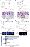
Figure 2. Clinical course of responders after initiation of IFN-γ.
Panels AD capture the courses of patients 1, 3, 4, and 6 (respectively), who achieved MRDneg CRs. Day 0 marks the start of IFN-γ therapy. The top plot depicts the kinetics of donor chimerism in unfractionated blood and BM and in the CD3+ and CD33+ fractions in blood. The lower plots show leukemia quantitation based on blast percentages by morphology and the percentages of CD34+ cells by flow cytometry (results from the clinical hematopathology and flow cytometry labs). The second row shows May-Grunwald-Giemsa–stained BM slides. The arrows indicate representative malignant cells (A and B). The third row shows representative BM morphology with CD34 staining (A and B) or FISH analyses of 9q34.13 NUP214 translocation using a “break apart” probe approach (C) or immunofluorescence for the X and Y chromosomes (D). (E) Timelines of the courses of all study participants. CR, complete remission; PR, partial remission; PD, progressive disease.
Figure 3
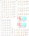
Figure 3. IFN-γ responsiveness of leukemia myeloblasts.
In vitro p-STAT1 quantitation of BM cells after in vitro culture with or without IFN-γ (1 or 50 ng/mL) for 15 minutes (A). Shown is anti–p-STAT1 staining gating on cells that were CD34+, CD13+CD11b, CD4+, CD8+, or CD19+. (B) Expression of HLA-ABC, HLA-DR, HLA-DP, ICAM-1, and PD-L1 on myeloblasts (see Supplemental Figure 1 for gating strategies) after culture for 48 hours without or with IFN-γ (1 ng/mL or 50 ng/mL). FMO (fluorescence minus 1) samples were cultured with 50 ng/mL IFN-γ. (C) BM samples from participants were collected between 2 and 14 days prior to and 48–60 hours after in vivo IFN-γ. Shown is expression of HLA-ABC, HLA-DR, HLA-DP, ICAM-1, and PD-L1 on myeloblasts. p-STAT1 and HLA-DR expression of an array of primary AML samples (n = 60) before and after in vitro IFN-γ stimulation (D and E). Shown is fold-induction with IFN-γ (y axis) for p-STAT1 (D) and HLA-DR (E) grouped by ELN classification (x axis). (F) Representative flow cytometry plots of p-STAT1 and IFN-γ–inducible molecules after in vitro culture with or without IFN-γ. Shown are data (from top to bottom) of samples that manifest IFN-γ induction of p-STAT1 and HLA-DR, only p-STAT1, or neither.
Figure 4
Figure 4. Serial measurements of CXCL10 (IP-10), IFN-γ, other cytokines, and the GVHD biomarkers ST2, Reg3α, and amphiregulin relative to clinical events.
(AD) Shown are data from patients 1–4, respectively.
Figure 5
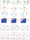
Figure 5. scRNA-Seq of BM samples collected before and after in vivo treatment with IFN-γ.
(A) Two-dimensional uniform manifold approximation and projection (UMAP) with cell lineage annotations for the combined pre–IFN-γ and post–IFN-γ samples. For patient 1, transcriptomes from the unfractionated and CD34-selected samples are also combined. (B) Donor and recipient origin of cells by SNP differences between donor and recipient in transcribed genes mapped onto the same UMAP as in A. Most early myeloid and erythroid cells were recipient in origin (consistent with relapsed MDS) whereas lymphoid cells and maturing monocytes were dominantly donor derived. (C) Pre– and post–IFN-γ samples are separately annotated. The overall distributions of major lineage clusters of the pre– and post–IFN-γ samples were similar. (D) SCPA. Shown are q values for changes in the listed pathways (higher values = greater changes in the pathway activity in the post–IFN-γ treatment sample). (E) Volcano plots of the expression of selected genes included in key gene sets in D for HSCs (patients 1 and 7) and myeloid progenitors (patient 5). Shown are Hallmark sets for “IFN-γ response” and Kyoto Encyclopedia of Genes and Genomes (KEGG) for genes involved in antigen processing and presentation. X axis shows average log2 fold-change in the post–IFN-γ treatment samples. Y axis shows –log10 of adjusted P values. (F) The dominant malignant lineage clusters shown in A were separately re-embedded. HSC-like cells were re-embedded for patients 1 and 7, and myeloid progenitor cells were re-embedded for patient 5. The cells in the clustering are depicted as UMAPs with the pre– and post–IFN-γ samples in separate plots. The representative expression levels of STAT1, HLA-A, CIITA, and HLA-DRA are shown in each patient. Ovals surround regions of differences in the re-embedded pre– and post–IFN-γ transcriptomes.

References

    1. Bleakley M, Riddell SR. Molecules and mechanisms of the graft-versus-leukaemia effect. Nat Rev Cancer. 2004;4(5):371–380. doi: 10.1038/nrc1365. - DOI - PubMed
    1. Alyea EP, et al. NCI First International Workshop on The Biology, Prevention and Treatment of Relapse after Allogeneic Hematopoietic Cell Transplantation: report from the committee on prevention of relapse following allogeneic cell transplantation for hematologic malignancies. Biol Blood Marrow Transplant. 2010;16(8):1037–1069. doi: 10.1016/j.bbmt.2010.05.005. - DOI - PMC - PubMed
    1. Bishop MR, et al. Introduction to the reports from the National Cancer Institute First International Workshop on the Biology, Prevention, and Treatment of Relapse after Allogeneic Hematopoietic Stem Cell Transplantation. Biol Blood Marrow Transplant. 2010;16(5):563–564. doi: 10.1016/j.bbmt.2010.02.025. - DOI - PMC - PubMed
    1. Wayne AS, et al. Proceedings from the National Cancer Institute’s Second International Workshop on the Biology, Prevention, and Treatment of Relapse after Hematopoietic Stem Cell Transplantation: introduction. Biol Blood Marrow Transplant. 2013;19(11):1534–1536. doi: 10.1016/j.bbmt.2013.08.016. - DOI - PMC - PubMed
    1. Horowitz M, et al. Epidemiology and biology of relapse after stem cell transplantation. Bone Marrow Transplant. 2018;53(11):1379–1389. doi: 10.1038/s41409-018-0171-z. - DOI - PMC - PubMed

Publication types

MeSH terms

Substances

Associated data