. 2025 Aug 14;96(9):908-911.
doi: 10.1136/jnnp-2024-335571.

Muscle MRI quantifies disease progression in amyotrophic lateral sclerosis

Affiliations

Muscle MRI quantifies disease progression in amyotrophic lateral sclerosis

Uros Klickovic et al. J Neurol Neurosurg Psychiatry. .

Abstract

Background and objectives: Quantitative and operator-independent biomarkers of disease progression are urgently needed in amyotrophic lateral sclerosis (ALS) research. We assess the potential of skeletal muscle MRI as a sensitive and reliable outcome measure for future ALS clinical trials.

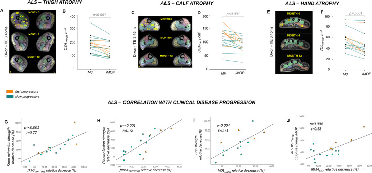
Methods: In this longitudinal cohort study, muscle MRI of head-neck, upper and lower limb regions, alongside clinical and functional assessments, were acquired at three time points over the individual maximum observation period (iMOP) of 1 year in 20 patients with ALS and 16 healthy controls. Quantitative MRI parameters cross-sectional area (CSA), volume (VOL), fat fraction, functional rest muscle area and water T2 (T2m) were correlated with changes in clinical disease severity (functional rating scales and myometry).

Results: Among 20 patients with ALS, 17 completed follow-up. Progressive muscle atrophy (CSA, VOL) was observed at hand (rs=0.66), head-neck (partial η²=0.47) and lower-limb level (thighs: η²=0.56, calves: η²=0.54) over iMOP. MRI changes correlated with leg muscle strength (knee extension: r=0.77; plantar flexion: r=0.78), hand grip strength (r=0.71) and functional rating scales (r=0.68).

Interpretation: Our findings demonstrate the effectiveness of muscle MRI as a sensitive neuroimaging biomarker of disease progression in ALS, highlighting its potential application in clinical trials.

Keywords: ALS; MRI; MUSCLE DISEASE; NEUROMUSCULAR.


Conflict of interest statement

Competing interests: None declared.

Figures

Figure 1
Figure 1. Muscle MRI detects progressive atrophy in lower limb and hand muscles in ALS and functional rest muscle area correlates with clinical disease progression. (A) Sample axial Dixon imaging (TE=3.45 ms) of thighs in patients with ALS at baseline (upper row), 6 months (middle row) and 12 months (lower row). (B) Mean values of overall cross-sectional area at thigh level (CSATHIGH) in patients with ALS (fast progressors: orange; slow progressors: green) at baseline (M0) and iMOP. CSATHIGH significantly decreased over the iMOP (T(1,16)=4.51, p<0.001). (C) Sample axial Dixon imaging (TE=3.45 ms) of calves in a patient with ALS at baseline (upper row), 6 months (middle row) and 12 months (lower row). (D) Mean values of overall CSA at calf level (CSACALF) in patients with ALS (fast progressors: orange; slow progressors: green) at baseline (M0) and iMOP. CSACALF significantly decreased over iMOP (T(1,16)=4.30, p<0.001). (E) Sample axial Dixon imaging (TE=3.45 ms) of the dominant hand of a patient with ALS at baseline (upper row), 6 months (middle row) and 12 months (lower row). (F) Mean values of hand volume (VOLHAND) in patients with ALS (fast progressors: orange; slow progressors: green) over time. VOLHAND significantly declined over the iMOP (test statistic=–122, p<0.001). (G) At thigh level, a correlation of the relative decrease of fRMAANT-TMC with the relative decrease of knee extension strength over the iMOP (r=0.77, p<0.001) was observed. (H) At calf level, the relative decrease of fRMATRICEPSSUR significantly correlated with the corresponding relative decrease of plantar flexion strength over iMOP (r=0.78, p<0.001). (I) At hand level, significant correlations were observed between the relative decrease in VOLHAND and the relative decrease of the grip strength over iMOP (r=0.71, p=0.004). (J) The relative change of fRMACALF showed a significant correlation with the absolute change of ALSFRS-RTOTAL score over the iMOP (r=0.68, p=0.004). ALS, amyotrophic lateral sclerosis; ALSFRS-R, ALS functional rating scale—revised; ALSFRS-RTOTAL, ALS functional rating scale total score; ALSFRS-RHAND-SS, ALS functional rating scale hand subscale score; CSA, cross-sectional area; CSATHIGH, overall cross-sectional area of thigh muscles; CSACALF, overall cross-sectional area of calf muscles; fRMAANT-TMC, muscle-specific functional remaining muscle area of anterior thigh muscle compartment; fRMATRICEPSSUR, muscle-specific functional remaining muscle area of triceps surae muscles; iMOP, individual maximum observation period; M0, baseline; M6, 6-month follow-up; M12, 12-month follow-up; VOLHAND, hand volume. Muscle abbreviations: AM, adductor magnus; BFP, biceps femoris posterior; G, gracilis; HT, hypothenar; IO, interossei; L, lumbricales; LG, lateral gastrocnemius; MG, medial gastrocnemius; PL, peroneus longus; RF, rectus femoris; S, sartorius; SM, semimembranosus; SO, soleus; ST, semitendinosus; TA, tibialis anterior; TH, thenar; TP, tibialis posterior: VL, vastus lateralis; VI, vastus intermedius; VM, vastus medialis.

References

    1. Morrow JM, Sinclair CDJ, Fischmann A, et al. MRI biomarker assessment of neuromuscular disease progression: a prospective observational cohort study. Lancet Neurol. 2016;15:65–77. doi: 10.1016/S1474-4422(15)00242-2. - DOI - PMC - PubMed
    1. Bonati U, Hafner P, Schädelin S, et al. Quantitative muscle MRI: A powerful surrogate outcome measure in Duchenne muscular dystrophy. Neuromuscul Disord. 2015;25:679–85. doi: 10.1016/j.nmd.2015.05.006. - DOI - PubMed
    1. Dahlqvist JR, Fornander F, de Stricker Borch J, et al. Disease progression and outcome measures in spinobulbar muscular atrophy. Ann Neurol. 2018;84:754–65. doi: 10.1002/ana.25345. - DOI - PubMed
    1. Klickovic U, Zampedri L, Sinclair CDJ, et al. Skeletal muscle MRI differentiates SBMA and ALS and correlates with disease severity. Neurology (ECronicon) 2019;93:e895–907. doi: 10.1212/WNL.0000000000008009. - DOI - PMC - PubMed
    1. Jenkins TM, Alix JJP, David C, et al. Imaging muscle as a potential biomarker of denervation in motor neuron disease. J Neurol Neurosurg Psychiatry. 2018;89:248–55. doi: 10.1136/jnnp-2017-316744. - DOI - PMC - PubMed

LinkOut - more resources