. 2025 Mar 25;44(1):107.
doi: 10.1186/s13046-025-03336-4.

Innovative dual-gene delivery platform using miR-124 and PD-1 via umbilical cord mesenchymal stem cells and exosome for glioblastoma therapy

Affiliations

Innovative dual-gene delivery platform using miR-124 and PD-1 via umbilical cord mesenchymal stem cells and exosome for glioblastoma therapy

Po-Fu Yueh et al. J Exp Clin Cancer Res. .

Abstract

Addressing the challenges of identifying suitable targets and effective delivery strategies is critical in pursuing therapeutic solutions for glioblastoma (GBM). This study focuses on the therapeutic potential of microRNA-124 (miR-124), known for its tumor-suppressing properties, by investigating its ability to target key oncogenic pathways in GBM. The results reveal that CDK4 and CDK6-cyclin-dependent kinases that promote cell cycle progression-are significantly overexpressed in GBM brain samples, underscoring their role in tumor proliferation and identifying them as critical targets for miR-124 intervention. However, delivering miRNA-based therapies remains a major obstacle due to the instability of RNA molecules and the difficulty in achieving targeted, efficient delivery. To address these issues, this research introduces an innovative, non-viral dual-gene delivery platform that utilizes umbilical cord mesenchymal stem cells (UMSCs) and their exosomes to transport miR-124 and programmed cell death protein-1 (PD-1). The efficacy of this dual-gene delivery system was validated using an orthotopic GBM model, which closely mimics the tumor microenvironment seen in patients. Experimental results demonstrate that the UMSC/miR-124-PD-1 complex and its exosomes successfully induce apoptosis in GBM cells, significantly inhibiting tumor growth. Notably, these treatments show minimal cytotoxic effects on normal glial cells, highlighting their safety and selectivity. Moreover, the study highlights the immunomodulatory properties of UMSC/miR-124-PD-1 and its exosomes, enhancing the activation of immune cells such as T cells and dendritic cells, while reducing immunosuppressive cells populations like regulatory T cells and myeloid-derived suppressor cells. The orchestrated dual-gene delivery system by UMSCs and exosomes showcased targeted tumor inhibition and positive immune modulation, emphasizing its potential as a promising therapeutic approach for GBM.

Keywords: CDK4/6; Gene therapy; Glioblastoma; Umbilical cord mesenchymal stem cells; miR-124.


Conflict of interest statement

Declarations. Studies involving animal subjects: The animal study conducted in this research was reviewed and approved by the Institutional Animal Care and Use Committee (IACUC) at China Medical University, Taichung, Taiwan (ID: CMU CMUIACUC-2019-042). Studies involving human subjects: Opensource data was used. Opensource data was used. The collection of brain tumor samples was approved by the Office of Human Research at Taipei Medical University (ID: N201901041). Inclusion of identifiable human data: There is no inclusion of identifiable human data in the manuscript. Consent for publication: All authors have agreed to the publication of the manuscript. Competing interests: The authors declare no competing interests.

Figures

Fig. 1
Fig. 1
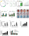
Identifying the Potential Role of CDK4 and CDK6 in GBM and Validating Them as Potential Target Factors Using miR-124. (A) RNA expression levels of CDK4 and CDK6 in GBM patients from Taiwan and (B) TCGA database assayed by RNA array and GEPIA platform. (C) Survival outcomes of GBM in different CDK4 and CDK6 expression levels. The dot line was represented as 95% confidence interval. (D) Protein and (E) RNA expression levels of CDK4 and CDK6 in glial cells and three different GBM cells. (F) The potential target region and the hypothesized regulation of miR-124 on both CDK4 and CDK6 assayed by TargetScan predicts tools. (G) The negative regulation between miR-124 and CDK4/CDK6 in glioma assayed by ENCORI Pan-Cancer Analysis Platform. (H) The miR-124 expression in glial cells and three different GBM cells assayed by q-PCR
Fig. 2
Fig. 2
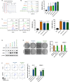
Characteristics of pre-miR-124 and PD-1 dual-gene-modified UMSCs and their capacity to induce apoptosis in GBM. (A) Construction of the pre-miR-124 and PD-1 dual-gene vector on PiggyBac transposon vectors. (B) Surface expression levels of PD-1 after electroporation with various vectors and selection of stable clones assayed by flow cytometry. (C) Protein expression of PD-1 in various vectors-transfected UMSCs assayed by Western blotting. (D) MiR-124 expression in various vectors-transfected UMSCs assayed by qPCR. (E-F) Proliferation and differentiation assays performed on various vectors-transfected UMSCs. (G-H) Early and late Annexin-V activation, (I) accumulation of BrdU, and (J) cleavage of caspase-3 in GBM cells co-cultured with different ratios of UMSC/miR-124-PD-1 assayed by flow cytometry. (K) Cell cycle regulation of GBM cells when co-cultured with different ratios of UMSC/miR-124-PD-1 assayed by flow cytometry
Fig. 3
Fig. 3
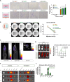
Characteristics of UMSC/miR-124-PD-1 derived exosome and their capacity to induce apoptosis in GBM. (A) Evaluation of exosome size derived from UMSC/miR-124-PD-1 by NTA. (B-D) Expression levels of CD9, CD63, CD81 on UMSC/miR-124-PD-1-derived exosomes assayed by flow cytometry and Western blotting. (E) Colony formation results in GBM cells co-cultured with exosomes derived from various vectors-transfected UMSCs. (F) Annexin-V activation in GBM cells co-cultured with exosomes derived from various vectors-transfected UMSCs assayed by flow cytometry
Fig. 4
Fig. 4
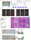
Infiltration, migration and bio-distribution characteristic of UMSC/miR-124-PD-1. The migration toward GBM cells is validated on various vectors-transfected UMSCs by (A) transwell assayed and (B) wound healing assay. (C) The infiltration of Ga-68 labeled UMSC/miR-124-PD-1 after ICA injection is detected by SPECT/CT scan. (D) Whole body distribution of DiR labeled UMSC/miR-124-PD-1 and its quantification signaling from brain area w/o tumor is determined by IVIS scan. (E) Ex vivo of DiR labeled UMSC/miR-124-PD-1 signaling from different organs at 2, 24 and 48 h is determined by IVIS scan in tumor bearing and normal mice
Fig. 5
Fig. 5
Therapeutic efficacy of UMSC/miR-124-PD-1 and its exosome on GBM-bearing mice. (A) Experimental flowchart for treatment evaluation in the GBM model using UMSCs transfected with various vectors. The (B) tumor volume assessed by MR T2-RARE scan and (C) survival outcome calculated using the Kaplan-Meier method for UMSCs transfected with various vectors are presented. (D) Whole-brain H&E stain from one represented mice is displayed. (E) Representative MR tumor images with three consecutive slices from each group of mice are displayed. (F) The body weight of each group of mice during treatment is presented. (G) Pathological evaluation of normal organs from each group of mice isolated on day 15 using H&E staining. (H) Biochemical analysis of liver function evaluated from mice serum on Day 15. (I-J) Protein expression of CDK4, CDK6, Ki-67 and cleaved caspase-3 in tumor tissue are assayed by IHC staining
Fig. 6
Fig. 6
Immunoregulation of UMSC/miR-124-PD-1 and its exosome on GBM-bearing mice. (A) CD4, CD8, CD86 induction signal, activation of (B) memory T cells, and (C) T cell proliferation in GBM co-cultured with UMSCs transfected with various vectors are assayed by flow cytometry. The accumulation of CD8+IFN-γ+ or CD8+IL-2+ CTL in mice (D) TDLN and (E) SP is assayed by flow cytometry. (F) CD11c+CD24+MHCII+ DCs in mice TDLN, (G) CD8+CD62L+CD44+ memory T cells in mice SP, and (H) CD11b+ CD86+ M1 in mice SP and BM are assayed by flow cytometry. (I) CD4+CD25+FOXP3+ Tregs in mice TDLN and SP, (J) CD11b+Gr-1+ MDSCs in mice SP and BM, and (K) CD11b+CD206+ M2 in mice SP and BM are assayed by flow cytometry. (L) CD8 and IFN-γ positive CTL cells are assayed by IF staining

References

    1. Wu W, Klockow JL, Zhang M, Lafortune F, Chang E, Jin L, Wu Y, Daldrup-Link HE. Glioblastoma multiforme (GBM): an overview of current therapies and mechanisms of resistance. Pharmacol Res. 2021;171:105780. - PMC - PubMed
    1. Nozhat Z, Heydarzadeh S, Shahriari-Khalaji M, Wang S, Iqbal MZ, Kong X. Advanced biomaterials for human glioblastoma multiforme (GBM) drug delivery. Biomaterials Sci. 2023;11(12):4094–131. - PubMed
    1. Abdul-Al M, Saeinasab M, Zare A, Barati M, Shakeri S, Keykhosravi E, Momeni-Moghaddam M, Najafzadeh M, Keshel SH, Farzi G, Sefat F. Application of biomaterials for glioblastoma treatment: promises, advances, and challenges. Mater Today Commun. 2022;33:104562.
    1. Yao Z, Jiang X, Yao H, Wu Y, Zhang F, Wang C, Qi C, Zhao C, Wu Z, Qi M, Zhang J, Cao X, Wang Z, Wu F, Yao C, Liu S, Ling S, Xia H. Efficiently targeted therapy of glioblastoma xenograft via multifunctional biomimetic nanodrugs. Biomaterials Res. 2022;26(1):71. - PMC - PubMed
    1. Xu H-Z, Li T-F, Ma Y, Li K, Zhang Q, Xu Y-H, Zhang Y-C, Zhao L, Chen X. Targeted photodynamic therapy of glioblastoma mediated by platelets with photo-controlled release property. Biomaterials. 2022;290:121833. - PubMed

MeSH terms