Review
. 2025 Mar 25;25(1):113.
doi: 10.1186/s12935-025-03754-6.

Migrasomes, critical players in intercellular communication

Affiliations
Review

Migrasomes, critical players in intercellular communication

Zhiyong Tan et al. Cancer Cell Int. .

Abstract

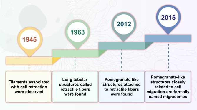
Migrasomes are a newly discovered type of extracellular vesicle (EV) formed during cell migration, playing a pivotal role in intercellular communication. These vesicles are generated by retracting fibers of migrating cells and encapsulate various molecules, such as proteins, lipids, and RNA, allowing the transfer of biochemical signals to neighboring cells. Current evidence suggests that migrasomes are involved in a wide range of physiological processes such as embryogenesis, angiogenesis, immune modulation, and mitochondrial quality control. Moreover, migrasomes are implicated in pathological conditions, including cancer metastasis, cardiovascular diseases, and viral infections. To fully understand their significance, it is critical to first explore the molecular mechanisms underlying their formation and function. Recent studies have shed light on the biogenesis, release, and biological properties of migrasomes, all of which are key to understanding their role in cell-to-cell communication. In this review, we provide an up-to-date summary of migrasome biogenesis, release, characterization, and their biological activities in intercellular communication, while also proposing potential new functions for these vesicles.

Keywords: Cell–cell communication; Extracellular vesicles; Migrasome; Physiological and pathological processes.


Conflict of interest statement

Declarations. Ethics approval and consent to participate: Not applicable. Consent for publication: Not applicable. Competing interests: The authors declare no competing interests.

Figures

Fig. 1
Fig. 1
Timeline of key discoveries leading to migrasome identification. In 1945, initial observations identified filaments associated with cell retraction [16]. By 1963, these were further characterized as long tubular structures known as retractile fibers [17]. In 2012, pomegranate-like structures were observed attached to these fibers [18]. The formal naming of these migratory, pomegranate-like structures as “migrasomes” occurred in 2015, linking them explicitly to cell migration [19]. This progression highlights the gradual unveiling of migrasomes’ unique morphology and their role in cellular dynamics
Fig. 2
Fig. 2
Extracellular vesicles (EVs) classifcation. This diagram provides a comprehensive overview of EV classification and biogenesis, emphasizing their size, cellular origin, and mechanisms of formation. EVs are categorized into small vesicles, including exomeres (<50 nm), exosomes (30–150 nm) derived from multivesicular bodies, and ectosomes (100–1000 nm) directly shed from the plasma membrane. The large vesicles comprise migrasomes (500–3000 nm) produced during cell migration, apoptotic bodies (1000–5000 nm) formed during apoptosis, and oncosomes (1000–10000 nm) associated with tumor cells. It emphasizes the distinct formation pathways for each EV type, including endosomal sorting, membrane budding, and cellular migration, demonstrating how these processes facilitate their release into the extracellular space
Fig. 3
Fig. 3
Stages of migrasome formation: nucleation, maturation, and expansion. Nucleation involves SMS2 catalyzing sphingomyelin formation at the cell membrane. Maturation is driven by PIP5K1A, PI(4)P, PI(4,5)P2, Rab35, and integrins, which promote membrane budding and protrusion development. Expansion involves the assembly of tetraspanin-enriched macrodomains (TEMs), extending the migrasome structure. Key molecular components like lipids, receptors, and integrins are crucial at each stage, emphasizing the pathways essential for migrasome development
Fig. 4
Fig. 4
Mechanisms of migrasome-mediated intercellular communication. The diagram depicts various biological roles associated with migrasomes, including the release of signaling molecules, shedding of cellular contents, and lateral transfer of materials between cells. Migrasomes facilitate communication and material exchange, influencing processes such as immune response, tissue repair, and development. The functions highlighted underscore the diverse and dynamic contributions of migrasomes to cellular and physiological processes, with some aspects remaining to be fully elucidated
Fig. 5
Fig. 5
Multifaceted physiological and pathological roles of migrasomes. Migrasomes are implicated in cardiovascular and cerebrovascular diseases (e.g., myocardial infarction, stroke), supporting cellular repair processes and influencing inflammatory pathways [67, 70]. They contribute to tumor progression in cancers such as gastric, liver, and glioma, and interact with viral pathogens like SARS-CoV-2, affecting immune responses and thrombosis [53, 56]. Additionally, migrasomes aid tissue regeneration, supporting fat and bone healing and providing early diagnostic potential for kidney injury, highlighting their pivotal role in intercellular communication and therapeutic applications [80]

References

    1. Lobos-Gonzalez L, Bustos R, Campos A, Silva V, Silva V, Jeldes E, Salomon C, Varas-Godoy M, Caceres-Verschae A, Duran E, et al. Exosomes released upon mitochondrial ASncmtRNA knockdown reduce tumorigenic properties of malignant breast cancer cells. Sci Rep. 2020;10(1):343. - PMC - PubMed
    1. Cocucci E, Meldolesi J. Ectosomes and exosomes: shedding the confusion between extracellular vesicles. Trends Cell Biol. 2015;25(6):364–72. - PubMed
    1. Kakarla R, Hur J, Kim YJ, Kim J, Chwae YJ. Apoptotic cell-derived exosomes: messages from dying cells. Exp Mol Med. 2020;52(1):1–6. - PMC - PubMed
    1. Minciacchi VR, Freeman MR, Di Vizio D. Extracellular vesicles in cancer: exosomes, microvesicles and the emerging role of large oncosomes. Semin Cell Dev Biol. 2015;40:41–51. - PMC - PubMed
    1. Jeppesen DK, Fenix AM, Franklin JL, Higginbotham JN, Zhang Q, Zimmerman LJ, Liebler DC, Ping J, Liu Q, Evans R, et al. Reassessment of exosome composition. Cell. 2019;177(2):428–e445418. - PMC - PubMed

LinkOut - more resources