. 2025 Mar 31;39(6):e70489.
doi: 10.1096/fj.202403301R.

In vivo synergistic enhancement of MIF-mediated inflammation in acute lung injury by the plant ortholog Arabidopsis MDL1

Affiliations

In vivo synergistic enhancement of MIF-mediated inflammation in acute lung injury by the plant ortholog Arabidopsis MDL1

Lukas Spiller et al. FASEB J. .

Abstract

Recent research has uncovered Arabidopsis thaliana proteins that are similar to the human inflammatory cytokine MIF. Plant MIF/D-dopachrome tautomerase (D-DT)-like proteins (MDLs) can interact with human MIF, yet the significance of these findings in living organisms has not been investigated. Given MIF's key role in acute respiratory distress syndrome promoting pulmonary inflammation, pathology, and leukocyte infiltration, here we set out to investigate the interplay between MIF and MDL1, one of three A. thaliana MIF orthologs, in an in vivo mouse model of MIF-induced acute lung injury (ALI). Human MIF and MDL1 were administered to C57BL/6 mice via inhalation, individually or in combination. Inhalation of MIF promoted various parameters of lung injury as evaluated by flow cytometry, immunofluorescence microscopy, RT-qPCR, and ELISA, while MDL1 inhalation alone had no effect. Intriguingly, combined treatment with MIF and MDL1 synergistically enhanced pulmonary infiltration of neutrophils and monocytic cells, accompanied by an upregulation of pro-inflammatory cytokine genes. Thus, the plant-derived MIF ortholog MDL1 potentiates MIF-induced inflammation in ALI. These data support the growing evidence of interactions between plant-derived compounds and human inflammatory mediators and illustrate how they may impact human health.

Keywords: MDL; MIF; acute lung injury; atypical chemokine; plant MIF.


Conflict of interest statement

J.B. is the inventor on patent applications related to anti‐MIF strategies. All other authors declare no competing interests.

Figures
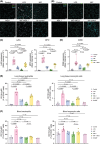
FIGURE 1
FIGURE 1
MDL1 amplifies MIF‐driven pulmonary infiltration of neutrophils and monocytic cells in an in vivo mouse model of acute lung injury (ALI). MDL1 amplified pulmonary leukocyte infiltration when co‐administered with human MIF, but not when applied alone. MIF, MDL1 or a combination of MIF and MDL1 (1:1) were applied to C57BL/6 mice via inhalation. After a 12 h incubation period, mice were sacrificed and pulmonary (lung tissue) neutrophil and monocytic cell levels were analyzed (A–D). Saline inhalation was used as negative control, LPS as positive control. To rule out species‐specific alterations, treatment with mouse MIF (MmMIF) was applied for comparison (E). (A) Representative fluorescent microscopy images as visualized by anti‐Ly6G (pink) and anti‐MPO (green) co‐staining. Cell nuclei were counterstained with DAPI (blue). Samples stained with secondary antibodies alone served as controls. Scale bar for all images is 100 μm (n = 10). (C) Quantification of the Ly6G+ and MPO+ mean fluorescence intensity according to (A); (data points from 10 randomly chosen images per mouse per experimental group). (B and D) Same as (A and C) except that infiltrated lung monocytic cells were visualized with anti‐CD68 antibody (green) and quantified. (E) Quantification of infiltrated cells by flow cytometry. Shown are neutrophils (left panel; CD45+CD11b+Ly6G+) and monocytic cells (right; CD45+CD11b+Ly6G) as percentage of CD45+ cells. (F) Corresponding flow cytometry from blood showing circulating neutrophils (left panel; CD45+CD11b+Ly6G+) and monocytic cells (right; CD45+CD11b+Ly6G). Values are shown as means ± SD with individual data points representing independent mice (n = 3–7). Statistical differences were analyzed using one‐way ANOVA with Tukey's post‐hoc test for E and F, with Dunnett's post‐hoc test for C (Ly6G) and Kruskal‐Wallis test with Dunn's post‐hoc test for C (MPO) and D. Statistical significance is indicated by p values. ns, not significant.
FIGURE 2
FIGURE 2
MDL1 amplifies MIF‐driven pro‐inflammatory cytokine levels in an in vivo mouse model of acute lung injury (ALI). (A and B) Heatmaps displaying (significant) changes in the mRNA expression of pro‐inflammatory cytokine genes using qRT‐PCR in C57BL/6 mouse lung tissue after nebulization with either human MIF, MDL 1 or the combination of MIF and MDL1 following an incubation period of 12 h. Saline inhalation is shown as negative control, inhalation of LPS served as positive control. Relative mRNA expression is shown relative to untreated controls with Rplp0 used as housekeeping gene. Shown is the fold change expression of (A) Tnf⍺; Ifnɣ; Il1β and (B) Ccl2; Il6; Cxcl2/Mip2 (n = 5–7, except Tnf⍺ / MIF experimental group n = 1). Statistical significance is indicated by p values (black p values indicate significance in comparison to control group, blue p values indicate significance between MIF and combination of MIF and MDL1 experimental groups). ns, not significant. (C) Upregulation of CCL2 as verified by ELISA from bronchoalveolar fluid (BALF). (D) Total protein count in BALF served as a surrogate parameter for lung injury and was measured photometrically using a BCA assay (n = 5–6). (E) Representative images of H&E‐stained mice lungs after stimulation as indicated. Scale bar for all images: 25 μm. Values are shown as means ± SD with individual datapoints representing independent mice. Statistical analysis was performed using one‐way ANOVA with Tukey's post‐hoc test for A (Tnf⍺ and Ifnɣ) and Dunnett's post‐hoc test for A (Il1β), B (Ccl2, Il6 and Cxcl2/Mip2), C and D. Statistical significance is indicated by p values. ns, not significant.

References

    1. Calandra T, Roger T. Macrophage migration inhibitory factor: a regulator of innate immunity. Nat Rev Immunol. 2003;3:791‐800. - PMC - PubMed
    1. Spiller L, Manjula R, Leissing F, et al. Plant MDL proteins synergize with the cytokine MIF at CXCR2 and CXCR4 receptors in human cells. Sci Signal. 2023;16:eadg2621. - PubMed
    1. Michelet C, Danchin EGJ, Jaouannet M, et al. Cross‐kingdom analysis of diversity, evolutionary history, and site selection within the eukaryotic macrophage migration inhibitory factor superfamily. Genes (Basel). 2019;10: 740. - PMC - PubMed
    1. Bernhagen J, Krohn R, Lue H, et al. MIF is a noncognate ligand of CXC chemokine receptors in inflammatory and atherogenic cell recruitment. Nat Med. 2007;13:587‐596. - PubMed
    1. Donnelly SC, Haslett C, Reid PT, et al. Regulatory role for macrophage migration inhibitory factor in acute respiratory distress syndrome. Nat Med. 1997;3:320‐323. doi:10.1038/nm0397-320 - DOI - PubMed

Substances

LinkOut - more resources