Review
. 2025 Mar 26;0(0):371-382.
doi: 10.1042/BST20240523.

Tetraspanins affect membrane structures and the trafficking of molecular partners: what impact on extracellular vesicles?

Affiliations
Review

Tetraspanins affect membrane structures and the trafficking of molecular partners: what impact on extracellular vesicles?

Eric Rubinstein et al. Biochem Soc Trans. .

Abstract

Tetraspanins are a family of 33 proteins in mammals believed to play a crucial role in the compartmentalization of various associated proteins within cells and membranes. Recent studies have elucidated the structure of several tetraspanin members, revealing that while the four transmembrane domains typically adopt a cone-shaped configuration in crystals, other conformations are also possible. This cone-shaped structure may explain why tetraspanins are often enriched in curved and tubular cellular structures, such as microvilli, tunneling nanotubes, retraction fibers, or at the site of virus budding, and may contribute to the formation or maintenance of these structures. Tetraspanins have also been detected on midbody remnants and migrasomes, as well as on extracellular vesicles (EVs), for which CD9, CD81, and CD63 are widely used as markers. Although their impact on certain membrane structures and their ability to regulate the function and trafficking of associated proteins would suggest a potential role of tetraspanins either in EV formation or in regulating their protein composition, or both, efforts to characterize these roles have been complicated by conflicting results. In line with the interaction of certain tetraspanins with cholesterol, two recent studies have suggested that the presence or organization of oxysterols and cholesterol in EVs may be regulated by Tspan6 and CD63, respectively, paving the way for further research on the influence of tetraspanins on the lipid composition of EVs.

Keywords: exosomes; extracellular vesicles; membrane dynamics; membrane proteins; microparticles; tetraspanins.


Conflict of interest statement

ER declares that there are no competing interests associated with the manuscript. PZ and CT are inventors on respectively 1 and 2 filed patents on the therapeutic use of EVs.

Figures

Figure 1
Figure 1. Structure of four tetraspanins.
The four transmembrane domains are in blue, the three structurally conserved helices in the large extracellular domain in red, and the variable region in green. The small extracellular domain is in gold. All structures were downloaded from PDB (www.rcsb.org). Top: the crystal structures of CD9 (PDB # 6K4J) and CD53 (PDB # 6WVG) and the cryo-EM structure of Tspan15 in association with ADAM10 (not shown). Bottom: the structure of CD81 (PDB # 5TCX) in crystals and that determined by cryo-EM (PDB # 7JIC) in which CD81 is in complex with CD19. Three different views of this structure are shown, including one showing CD19. Note that in this structure, CD81 does not show an intramembrane cavity and that the LEL extends from the plane of the membrane.
Figure 2
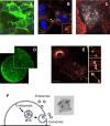
Figure 2. Examples of tetraspanin localization on remarkable membrane structures.
(A): Localization of GFP-tagged CD9 on tunneling nanotubes in SH-SY5Y cells (image kindly provided by Dr. C Brou (see: [28]). (B): Localization of CD9 (red) and CD81 (green) on midbody remnants in HeLa cells, similarly to what has been described in [29]. (C): CD9 (red), detected by super-resolution microscopy concentrates in areas in which the HIV protein GAG (white) accumulates and induces membrane bending (image kindly provided by Dr. PE Milhiet; see: [30]). (D): CD9 expression in oocytes. The punctuated pattern reflects its preferential localization on microvilli as shown by [31]. (E): GFP-tagged Tspan4 localizes on retraction fibers and migrasomes. (image reproduced from [32]; https://doi.org/10.1016/j.cellin.2021.100003). (F): Small extracellular vesicles are released by either direct budding from the plasma membrane (ectosomes, enriched in CD9 and CD81) or following the fusion of multivesicular endosome resulting in the release of their intraluminal vesicles (exosomes, enriched in CD63). A labeling of CD63 on extracellular vesicle analyzed by electronic microscopy is also shown (image reproduced from [33], https://doi.org/10.3390/antib9030029).

References

    1. Huang S., Yuan S., Dong M., Su J., Yu C., Shen Y., et al. The phylogenetic analysis of tetraspanins projects the evolution of cell-cell interactions from unicellular to multicellular organisms. Genomics. 2005;86:674–684. doi: 10.1016/j.ygeno.2005.08.004. - DOI - PubMed
    1. Garcia-España A., Chung P.J., Sarkar I.N., Stiner E., Sun T.T., Desalle R. Appearance of new tetraspanin genes during vertebrate evolution. Genomics. 2008;91:326–334. doi: 10.1016/j.ygeno.2007.12.005. - DOI - PubMed
    1. Hemler M.E. Tetraspanin functions and associated microdomains. Nat. Rev. Mol. Cell Biol. 2005;6:801–811. doi: 10.1038/nrm1736. - DOI - PubMed
    1. Charrin S., Le Naour F., Silvie O., Milhiet P.E., Boucheix C., Rubinstein E. Lateral organization of membrane proteins: tetraspanins spin their web. Biochem. J. 2009;420:133–154. doi: 10.1042/BJ20082422. - DOI - PubMed
    1. Charrin S., Jouannet S., Boucheix C., Rubinstein E. Tetraspanins at a glance. J. Cell. Sci. 2014;127:3641–3648. doi: 10.1242/jcs.154906. - DOI - PubMed

Publication types

LinkOut - more resources