. 2025 Apr 9;27(15):7605-7610.
doi: 10.1039/d5cp00410a.

Color variation in radio-luminescence of P-dots doped with thermally activated delayed fluorescence molecules

Affiliations

Color variation in radio-luminescence of P-dots doped with thermally activated delayed fluorescence molecules

Zheming Su et al. Phys Chem Chem Phys. .

Abstract

Thermally activated delayed fluorescence (TADF) materials possess exceptional photophysical properties. Organic scintillators utilizing TADF materials have shown great promise for applications requiring efficient radio-luminescence, owing to their high quantum efficiency and tunable emission properties. Previous studies demonstrated that polymer dots (P-dots) doped with TADF materials exhibit radio-luminescence under hard X-ray and electron beam excitation. However, the TADF materials used in these experiments were limited to limited color options, restricting their utility and hindering the exploration of multicolor radio-luminescence necessary for advanced applications. In this study, we successfully achieved multicolor radio-luminescence-blue, yellow, and red-by developing P-dots doped with TADF materials that emit across the visible spectrum. This breakthrough was demonstrated under excitation by hard X-rays, gamma rays, and electron beams. The ability to realize multicolor radio-luminescence is crucial, as it enables enhanced spatial and spectral resolution, which is vital for applications such as high-precision bio-imaging and multimodal sensing.


Conflict of interest statement

There are no conflicts to declare.

Figures

Fig. 1
Fig. 1. (a) Conceptual diagram of TADF P-dots showing radio-luminescence exhibiting various colors. (b) Chemical structures of TADF molecules used in this study. (c) Photographs of P-dots (1), P-dots (2) and P-dots (3) under room light (top) and ultra-violet light (365 nm) (bottom). (d) Normalized photoluminescence spectra of P-dots (1), P-dots (2) and P-dots (3) excited at 355 nm.
Fig. 2
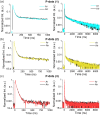
Fig. 2. Time-resolved photoluminescence decay curves for P-dots (1) (a), P-dots (2) (b), and P-dots (3) (c) in different timescales (left: 0–1000 ns, right: 0–9000 ns). Measurements taken under argon atmosphere are shown in color (sky blue, yellow, and red), while those taken in the presence of molecular oxygen are shown in black.
Fig. 3
Fig. 3. Decay profiles of transient absorption for P-dots (1) (a) at 550 nm, P-dots (2) (b) at 500 nm, and P-dots (3) (c) at 430 nm excited at 355 nm. Measurements taken under argon atmosphere are shown in color (sky blue, yellow, and red), while those taken in the presence of oxygen are shown in black.
Fig. 4
Fig. 4. Time dependence of scintillation intensity by gamma ray excitation for P-dots (1) (a), P-dots (2) (b), and P-dots (3) (c).
Fig. 5
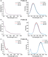
Fig. 5. Stability test upon gamma-ray irradiation. UV (Left) and emission spectra excited at 355 nm (right), before and after gamma ray irradiation for (a) P-dots (1), (b) P-dots (2) and (c) P-dots (3) at a dose rate of 17.45 Gy h−1 from a distance of 1 m for 30 or 60 min.
Fig. 6
Fig. 6. (a) Hard X-ray excited radioluminescence on TADF P-dots film observed by a CCD camera, with the X-ray beam directed toward the P-dots films positioned approximately 30 cm below the X-ray aperture. All scale bars are 10 mm. (b) and (c) Scintillation emission spectra for P-dots (1) (b) and P-dots (2) (c), where each film sample was measured three times with an exposure time of 5 seconds per measurement for averaging. X-ray parameters for both experiments: 60 kVp and 40 mA, unfiltered for P-dots (1) (b) and P-dots (2) (c).
Fig. 7
Fig. 7. (a) Photographs of films containing P-dots under room light (top) and under ultraviolet light (365 nm). (b) Photoluminescence spectra of films containing P-dots excited at 355 nm.
Fig. 8
Fig. 8. Radio luminescence of P-dots observed by electron beam irradiation. (a) Photographs for P-dots (1), P-dots (2), and P-dots (3). (b) The experimental setup photograph. (c) Radio-luminescence spectra for P-dots (1) (sky blue), P-dots (2) (yellow), and P-dots (3) (red).

References

    1. Shi Y.-Z. Wu H. Wang K. Yu J. Ou X.-M. Zhang X.-H. Chem. Sci. 2022;13:3625–3651. doi: 10.1039/D1SC07180G. - DOI - PMC - PubMed
    1. Bryden M. A. Zysman-Colman E. Chem. Soc. Rev. 2021;50:7587–7680. doi: 10.1039/D1CS00198A. - DOI - PubMed
    1. Li T. Yang D. Zhai L. Wang S. Zhao B. Fu N. Wang L. Tao Y. Huang W. Adv. Sci. 2017;4:1600166. doi: 10.1002/advs.201600166. - DOI - PMC - PubMed
    1. Osakada Y. Pratx G. Hanson L. Solomon P. E. Xing L. Cui B. Chem. Commun. 2013;49:4319–4321. doi: 10.1039/C2CC37169C. - DOI - PubMed
    1. Liu Z. Su Z. Asanuma D. Tojo S. Yamaji M. Fujitsuka M. Osakada Y. Photochem. Photobiol. Sci. 2024;23:329–338. doi: 10.1007/s43630-023-00520-3. - DOI - PubMed

LinkOut - more resources