. 2025 Mar;21(3):e70087.
doi: 10.1002/alz.70087.

Pathways underlying selective neuronal vulnerability in Alzheimer's disease: Contrasting the vulnerable locus coeruleus to the resilient substantia nigra

Affiliations

Pathways underlying selective neuronal vulnerability in Alzheimer's disease: Contrasting the vulnerable locus coeruleus to the resilient substantia nigra

Alexander J Ehrenberg et al. Alzheimers Dement. 2025 Mar.

Abstract

Introduction: Alzheimer's disease (AD) selectively affects certain brain regions, yet the mechanisms of selective vulnerability remain poorly understood. The neuromodulatory subcortical system, which includes nuclei exhibiting a range of vulnerability and resilience to AD-type degeneration, presents a framework for uncovering these mechanisms.

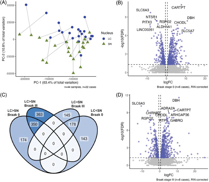
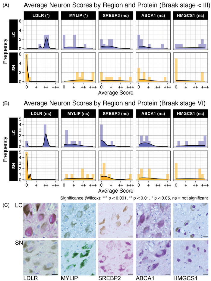
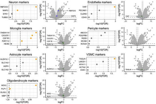
Methods: We leveraged transcriptomics and immunohistochemistry in paired samples from human post mortem tissue representing a vulnerable and resilient region-the locus coeruleus (LC) and substantia nigra (SN). These regions have comparable anatomical features but distinct vulnerability to AD.

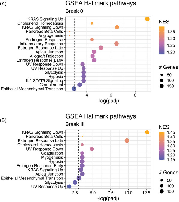
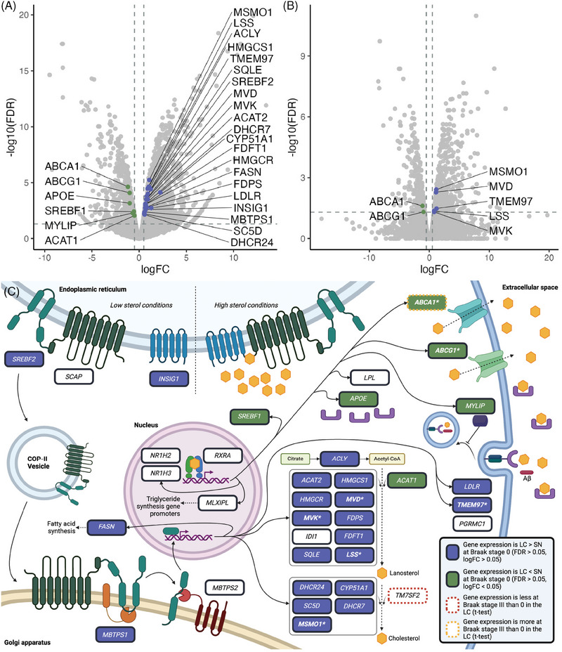
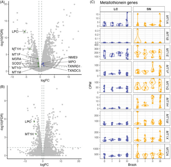
Results: We identified significant differences in cholesterol homeostasis, antioxidant pathways, KRAS signaling, and estrogen signaling at a bulk transcriptomic level. Notably, evidence of sigma-2 receptor upregulation was detected in the LC.

Discussion: Our findings highlight pathways differentiating the LC and SN, potentially explaining the LC's selective vulnerability in AD. Such pathways offer potential targets of disease-modifying therapies for AD.

Highlights: Intraindividual comparative RNAseq was used to study selective vulnerability. Metallothionein genes are significantly enriched in the substantia nigra. Cholesterol homeostatic genes are significantly enriched in the locus coeruleus. The locus coeruleus is likely more susceptible to toxic amyloid beta oligomers.

Keywords: Alzheimer's disease; autopsy; catecholamines; dopamine; human; locus coeruleus; neuropathology; noradrenaline; substantia nigra; tauopathy; transcriptomics.


Conflict of interest statement

A.J.E. has no relevant competing financial interests with this manuscript. A.J.E. serves as an unpaid steering committee member for the Genomic Answers for Children's Health Alliance at Leavitt Partners and as an unpaid executive committee member for the Neuromodulatory Subcortical System's Professional Interests Area of the International Society to Advance Alzheimer's Research and Treatment. S.T. has no relevant competing interests with this manuscript, but is co‐founder of Luxa Biotech developing an RPE therapy for AMD and has patents related to RPE cell therapy: Retinal pigment epithelial stem cells, Patent number: 8481313; Methods of treating a retinal disease by Retinal pigment epithelial stem cells, Patent number: 10034916; S.T. has advised BlueRock Therapeutics, Vita Therapeutics, and SANA Biotechnology. All other authors have no declared conflicts of interest to declare. Author disclosures are available in the Supporting Information.

Figures

FIGURE 1
FIGURE 1
A, The matched LC and SN samples (n = 22) segregate along PC‐2 in a PCA of gene expression for all matched samples. Connecting lines between points denote the case that both samples were collected from. B, There were 845 DEGs between the LC and SN in Braak stage 0 matched samples (n = 6, SN = baseline) in the RIN‐corrected and 1036 DEGs in the Braak stage III matched samples (n = 8, SN = baseline) in the RIN‐corrected model (D). C, Of the 524 genes more expressed in the LC than the SN in Braak 0, 350 remained more expressed in the LC in Braak III. Of the 321 genes more expressed in the SN than the LC in Braak 0, 178 remained more expressed in the SN than the LC in Braak III. DEG, differentially expressed gene; LC, locus coeruleus; logFC, log fold change; PC‐2, principal component 2; PCA, principal component analysis; RIN, RNA integrity number; SN, substantia nigra
FIGURE 2
FIGURE 2
A, In Braak stage 0 cases, the RIN‐corrected DEGs between the LC and SN showed statistically significant (P adj < 0.05) enrichment for 16 Hallmark pathways. B, In Braak stage III cases, statistically significant enrichment was found for 12 Hallmark pathways. DEG, differentially expressed gene; GSEA, gene set enrichment analysis; LC, locus coeruleus; RIN, RNA integrity number; SN, substantia nigra
FIGURE 3
FIGURE 3
A list of 37 cholesterol‐regulating or producing genes was examined among Braak stage 0 cases. A, In the uncorrected model of Braak stage 0 cases (SN = baseline), six genes (left‐justified text, green dots) were less expressed in LC than in SN (logFC < −0.5; FDR < 0.05) and 21 genes (right‐justified text, blue dots) were more expressed in LC than in SN (logFC > 0.5; FDR < 0.05). B, In the RIN‐corrected model at Braak stage 0 (SN = baseline), two genes were less expressed in LC than in SN and five were more expressed in LC than in SN. C, The genes more expressed in LC at Braak stage 0 in the uncorrected model are mostly involved in SREBP‐mediated production and influx of cholesterol. The genes less expressed in the LC at Braak stage 0 in the uncorrected model are mostly involved in LXR/RXR‐mediated transport and efflux of cholesterol. FDR, false discovery rate; LC, locus coeruleus; logFC, log fold change; RIN, RNA integrity number; SN, substantia nigra
FIGURE 4
FIGURE 4
Five differentially expressed genes from cholesterol pathways were evaluated at the protein level using IHC and semi‐quantitative assessment in Braak stage 0 to II (A) and Braak stage VI (B) cases. Neurons within a 500 × 500  µm region of interest were scored from 0 to +++. A numeric value was assigned to the scores (0 = 0, + to 1, ++ to 2, and +++ to 3) and each nucleus for each case received an average score. Paired Wilcoxon rank‐sum tests without exact matching were used to test for statistically significant differences between the two nuclei. All markers evaluated had neuronal expression in at least one nucleus. Representative images (C) from a 67‐year‐old female at Braak stage 0 (Case #11, Table 2) are provided. Scale bars: 20 µm. IHC, Immunohistochemistry; LC, locus coeruleus; SN, substantia nigra
FIGURE 5
FIGURE 5
Genes encoding markers of microglia, astrocytes, oligodendrocytes, endothelial cells, pericytes, vascular smooth muscle cells, and neurons were examined in the uncorrected Braak stage 0 differential expression results in LC and SN (SN = baseline). Cutoffs for statistical significance include FDR of 0.05 and a logFC of 0.5. Genes passing these thresholds are denoted by yellow coloring in the dot plots and labeling in the volcano plots. FDR, false discovery rate; LC, locus coeruleus; logFC, log fold change; SN, substantia nigra; VSMC, vascular smooth muscle cells
FIGURE 6
FIGURE 6
Differential expression of genes in LC and SN (logFC > 0 indicates greater expression of a gene in the LC). Cutoffs for significance are FDR < 0.05 and logFC > 0.5 or < −0.5. A–B, Significantly differentially expressed genes representing antioxidant enzymes or antioxidant activity selected from Gelain et al. are labeled for analyses of cases at Braak stage 0 in uncorrected (A) and RIN‐corrected (B) models. C, Metallothionein genes listed in Gelain et al. are expressed in both the LC and SN, with some more expressed in the SN relative to the LC. SN = baseline. FDR, false discovery rate; LC, locus coeruleus; logFC, log fold change; RIN, RNA integrity number; SN, substantia nigra

Similar articles

References

    1. Rub U, Stratmann K, Heinsen H, et al. The brainstem tau cytoskeletal pathology of Alzheimer's disease: a brief historical overview and description of its anatomical distribution pattern, evolutional features, pathogenetic and clinical relevance. Curr Alzheimer Res. 2016;13(10):1178‐1197. doi:10.2174/1567205013666160606100509 - DOI - PubMed
    1. Stratmann K, Heinsen H, Korf HW, et al. Precortical phase of Alzheimer's disease (AD)‐Related tau cytoskeletal pathology. Brain Pathol. 2016;26(3):371‐386. doi:10.1111/bpa.12289 - DOI - PMC - PubMed
    1. Attems J, Thomas A, Jellinger K. Correlations between cortical and subcortical tau pathology. Neuropath Appl Neuro. 2012;38(6):582‐590. doi:10.1111/j.1365-2990.2011.01244.x - DOI - PubMed
    1. Ehrenberg AJ, Kelberman MA, Liu KY, et al. Priorities for research on neuromodulatory subcortical systems in Alzheimer's disease: position paper from the NSS PIA of ISTAART. Alzheimers Dement. 2023;19(5):2182‐2196. doi:10.1002/alz.12937 - DOI - PMC - PubMed
    1. Ehrenberg AJ, Nguy AK, Theofilas P, et al. Quantifying the accretion of hyperphosphorylated tau in the locus coeruleus and dorsal raphe nucleus: the pathological building blocks of early Alzheimer's disease. Neuropathol Appl Neurobiol. 2017;43(5):393‐408. doi:10.1111/nan.12387 - DOI - PMC - PubMed