. 2025 Mar 26:10.1002/dvdy.70014.
doi: 10.1002/dvdy.70014. Online ahead of print.

Sox9 in the second heart field and the development of the outflow tract; implications for cardiac septation and valve formation

Affiliations

Sox9 in the second heart field and the development of the outflow tract; implications for cardiac septation and valve formation

Jenna R Drummond et al. Dev Dyn. .

Abstract

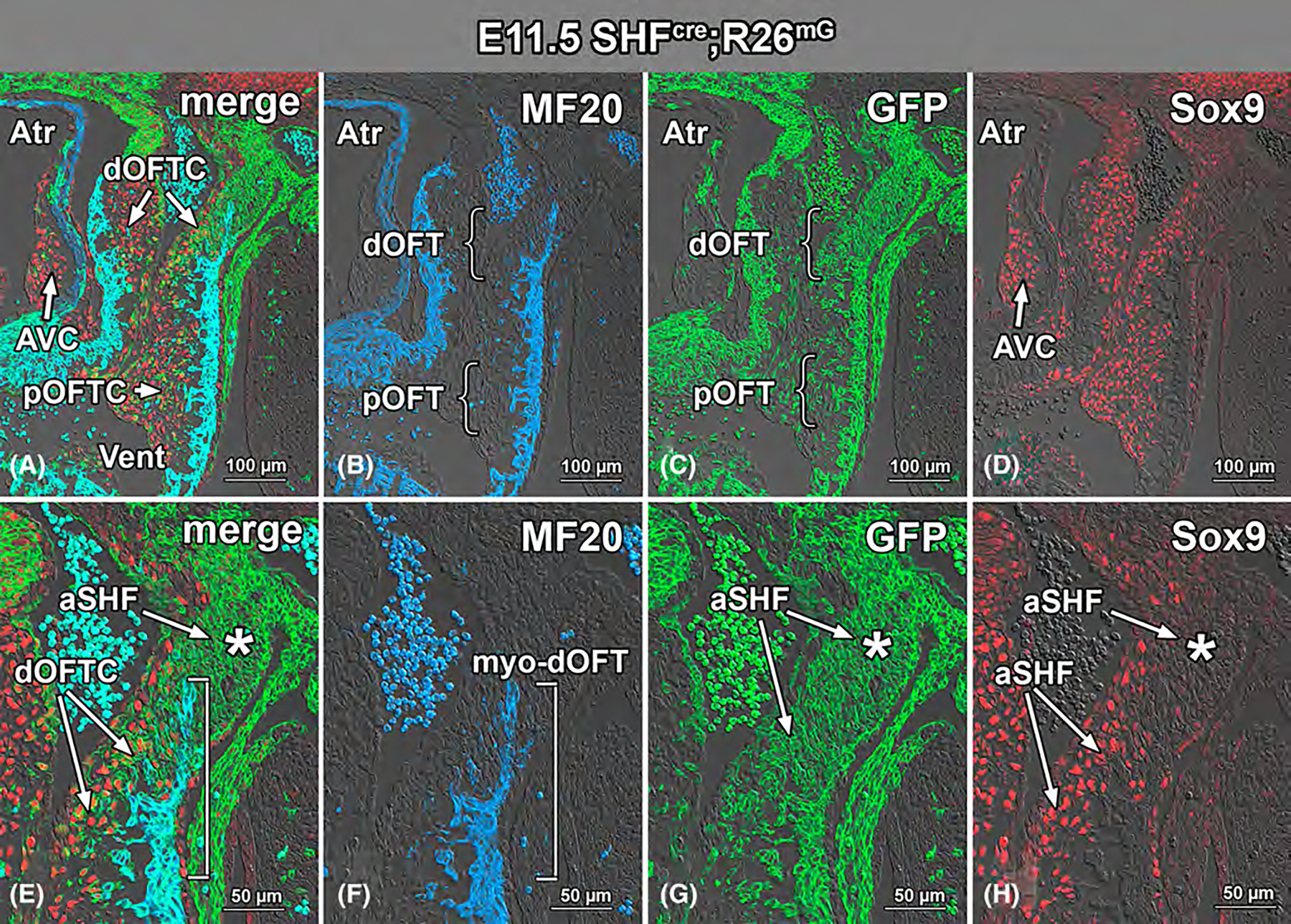
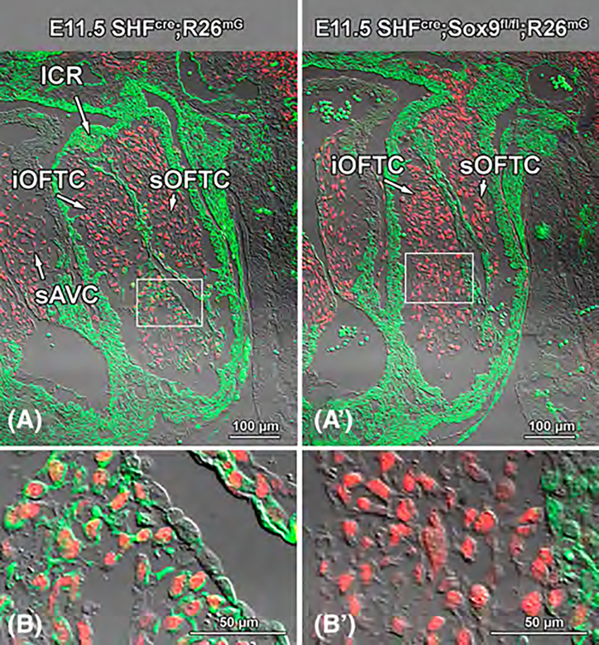
Background: Previously, we explored the role of Sox9 in the second heart field (SHF) in atrioventricular septation. For that study, we created a SHF-specific Sox9 knockout mouse. In addition to the presence of primary atrial septal defects in half of the offspring, we found that virtually all specimens also developed a ventricular septal defect. Histological analysis suggested that the ventricular septal defects resulted from developmental perturbation of the mesenchymal structures within the outflow tract. In the current study, we investigated the role of Sox9 in the SHF in the development of these tissues.

Results: Sox9 is expressed in all mesenchymal cell populations in the developing outflow tract, including a cohort of endocardial-derived cells that originate from the SHF-derived endocardium. SHF-specific deletion of Sox9 inhibits the formation of this cell population and ultimately leads to truncation of the mesenchymal outlet septum. This prevents complete fusion of this outlet septum with the atrioventricular mesenchymal complex, resulting in ventricular septal defects.

Conclusions: In combination with our first paper on the role of Sox9 in atrioventricular septation, data presented in this study demonstrate that Sox9 expression in the SHF is of critical importance for the proper formation of the septal structures in the developing heart.

Keywords: cell lineages; defects; development; heart; sox transcription factors.


Conflict of interest statement

CONFLICT OF INTEREST STATEMENT

Authors have no conflict of interest to disclose. All experiments were approved by the MUSC Institutional Animal Care and Use Committee and complied with all federal and institutional guidelines.

Figures

FIGURE 1
FIGURE 1
SOX9 expression in the outflow tract at E9.25. Sections of a sagitally sectioned E9.25 SHFcre;R26mG embryo were stained for GFP (green in C, G), MF20 (blue in B, F), and SOX9 (red in D, H). In panel A, the images in B, C, and D are merged, in panel E, the images in F, G, and H are merged. The staining for GFP in C, G shows that the SHF contributes to the myocardium RV and OFT (C), while SHF-derived cells are not observed in atrial and LV myocardium. The SOX9 staining in D shows limited expression of SOX9 in some endocardial cells in the OFT. A, atrium; AVC, atrioventricular cushions; CJ, cardiac jelly; endo, endocardium; LV, left ventricle; OFT, outflow tract; PE, proepicardium; pha-mes, pharyngeal mesenchyme; RV, right ventricle; SV, sinus venosus.
FIGURE 2
FIGURE 2
SOX9 expression in the outflow tract at E10. Section of a transversally sectioned Wnt1Cre;R26mG embryo at E10 stained for GFP (green; A, B), SOX9 (red; A, B), and MF20 (blue; A, B). Panel B is an enlargement and shows the upper part of the OFT in more detail. The panels show the migration of the GFP-expressing and SOX9-positive cardiac neural crest cells (CNCCs) into the distal part of the OFT. Note the expression of SOX9 in some endocardial cells. APS, aortopulmonary septum; CNCCs, cardiac neural crest cells; endo, endocardium; iOFTC, inferior outflow tract cushion; OFT, outflow tract; RA, right atrium; RV, right ventricle; sOFTC, superior outflow tract cushion.
FIGURE 3
FIGURE 3
The development of the intercalated ridges at E10.5. This figure shows the distal end of the OFT of a sagitally sectioned SHFCre;R26mG embryo at E10.5 at the level of a developing ICR. Panels A and D are low magnifications allowing easier orientation, while panels B, C, E, and F show the individual channels at higher magnification. The section was quadruple immunofluorescently stained for GFP (teal; A, B), Isl1 (green; A, C), MF20 (blue; A, D, E) and SOX9 (red; A, F). In panel A, all the channels are merged. The figure shows that the GFP-positive, Isl1-expressing ICR is emerging from a section of the OFT wall that is not expressing the myocardial marker MF20. Note that the developing ICR itself is not expressing SOX9 (A, F). The SOX9-expressing cells sandwiched between the endocardium and myocardium are CNCCs (A, F). CNCCs, cardiac neural crest cells; endo, endocardium; ICR, intercalated ridge; myo, myocardium; OFT, outflow tract.
FIGURE 4
FIGURE 4
SOX9 expression in the cardiac neural crest and endocardial-derived cells at E11.5. Panels A and B show a section of a Wnt1Cre;R26mG embryo at E11.5 (B is enlargement of A) stained for GFP (green), SOX9 (red), and MF20 (blue) illustrating the contribution of the SOX9-expressing CNCCs to the developing OFT at this stage. Panels C and D show a section of a Tie2Cre;R26mG embryo at E11.5 (D is enlargement of C) stained for GFP (green), SOX9 (red), and MF20 (blue) showing that ENDCs form a rather small percentage of the total of mesenchymal cells in the developing cushions of the OFT. AVCs, atrioventricular cushions; CNCCs, cardiac neural crest-derived cells; endo, endocardium; ENDCs, endocardial-derived cells; OFT, outflow tract; RV, right ventricle.
FIGURE 5
FIGURE 5
SOX9 expression in the anterior SHF-derived cells in the outflow tract at E10.5 and E11.5. Sections of SHFCre;R26mG embryos at E10.5 (A–C) and E11.5 (D–F) were stained for GFP (green; A–F) and SOX9 (red; A, C, D, F). Panel A (enlarged in C) shows the emerging population of aSHF-derived ENDCs at E10.5, while the image in D (enlarged in F) shows that at E11.5, the population of aSHF/ENDCs has expanded. The GFP-negative/SOX9-positive mesenchymal cells in A and D represent the population of CNCCs in the OFT cushions. aSHF/ENDCs, endocardial-derived cells with aSHF origin; CNCCs, cardiac neural crest-derived cells; dOFT, distal OFT; pOFT, proximal OFT.
FIGURE 6
FIGURE 6
Incorporation of aSHF cells into the distal part of the OFT cushions. This figure shows the OFT of an E11.5 SHFCre;R26mG embryo in sagittal orientation. Panels A–D are at low magnification, whereas E–H show the most distal part of the OFT in more detail. The tissue was stained for GFP (green; A, C, E, G), for the myocardial marker MF20 (blue; A, B, E, F), and for SOX9 (red; A, D, E, H). Combined, the panels illustrate how a population of Sox9-positive aSHF cells becomes incorporated into the upper part of the OFT cushion tissues as the OFT develops. Note that the majority of aSHF cells within the dOFTC express Sox9, whereas the majority of cells located distal to the myocardial cuff of the OFT do not (asterisk in G, H). aSHF, anterior second heart field; Atr, atrium; AVC, atrioventricular cushion; dOFT, distal outflow tract; dOFTC, distal outflow tract cushion; pOFT, proximal outflow tract; pOFTC, proximal outflow tract cushion; myo-dOFT, myocardial part of distal outflow tract.
FIGURE 7
FIGURE 7
The contribution of the SHF and expression of SOX9 and the formation of the semilunar valves at E12.5. This figure illustrates the formation of the pulmonary (A–D) and aortic valves (E–H) in a SHFCre;R26mG embryo at E12.5. A and E are merged images of the individual staining patterns for GFP (green; A, B, E, F), SOX9 (red; A, C, E, G), and smooth muscle actin (blue; A, D, E, H). Note the GFP and SOX9 staining of the intercalated ridges (B, C, F, G). Panel I is a schematic representation illustrating how the individual leaflet precursors from OFT cushions and intercalated ridges contribute to the respective leaflets of the semilunar valves. aICR, aortic ICR; pICR, pulmonary ICR; OFTc, OFT cushion; iOFTC, inferior OFT cushion; sOFTS, superior OFT cushion; al-PuV, anterior leaflet of pulmonary valve; pl-AoV, posterior leaflet of aortic valve; rl-PuV, right lateral leaflet of pulmonary valve; rl-AoV, right lateral leaflet of aortic valve; llPuV, left lateral leaflet of pulmonary valve; ll-AoV, left lateral leaflet of aortic valve.
FIGURE 8
FIGURE 8
Regionalized distal-to-proximal contribution of the aSHF, cardiac neural crest, and endocardium to the mesenchyme of the outflow tract at E12.5. This figure illustrates how aSHF, cardiac neural crest, and endocardium contribute to the mesenchyme in the OFT cushions at E12.5. GFP staining (green) represent the Cre-mediated GFP expression in SHFCre;R26mG (A, A′, A″), WntCre;R26mG (B, B′, B″), and Tie2Cre;R26mG (C, C′, C″) specimens. MF20 staining (blue) marks the myocardial structures, and the expression of SOX9 is visualized in red. Note the significant contribution of the aSHF to the most distal part of the OFT cushions (A, A’), the contribution of the CNCCs to the middle portion of the OFT cushions (B, B″), and the localization of the endocardial-derived cells in the proximal part of the OFT cushions (C, C″). All mesenchymal cells in the cushions express SOX9. dOFTC, distal OFT cushion; pOFTC, proximal OFT cushion.
FIGURE 9
FIGURE 9
Visualization and quantification of the contribution of SHF, cardiac neural crest, and endocardium to the OFT cushionderived semilunar valve leaflets at E12.5. For this experiment, lineage-specific Cre mice were used in combination with the ROSA26nT/nG nuclear reporter mouse. Panels A, A′ show a section of a SHFCre;R26nG embryo at E12.5, panels B, B′ show a section from a WntCre;R26nG embryo at E12.5, and C, C′ from a Tie2Cre;R26nG at the same stage. GFP staining (green) represents the Cre-mediated GFP expression, MF20 staining (blue; A-C) delineates the myocardial tissues, and SOX9 expressing cells are shown in red (A, B, C). This figure illustrates the significant contribution of the aSHF to the developing leaflets of the semilunar valves (A, A′) and the very limited contribution of CNCCs (B, B′) and (C, C′) to these valves. Panel E graphically illustrates the relative contribution of each cell lineage to the respective OFT cushion derived developing semilunar valve leaflets (see also text in body of manuscript). Panel D is for additional illustration of location of developing leaflets (see also Figure 7I). al-PuV, anterior leaflet of pulmonary valve; pl-AoV, posterior leaflet of aortic valve; rl-PuV, right lateral leaflet of pulmonary valve; rl-AoV, right lateral leaflet of aortic valve; ll-PuV, left lateral leaflet of pulmonary valve; ll-AoV, left lateral leaflet of aortic valve.
FIGURE 10
FIGURE 10
Deletion of SOX9 from the SHF results in ventricular septal defects. Hematoxylin/eosin-stained sections of control hearts (A, C) and SHFCre;SOX9fl/fl (B, D) at E15 (A, B) and E18.5 (C, D) demonstrate the ventricular septal defect in the second heart field-specific SOX9 knockout mouse. AoV, aortic valve; IVS, interventricular septum; LV, left ventricle; RV, right ventricle; VSD, ventricular septal defect.
FIGURE 11
FIGURE 11
Deletion of SOX9 from the SHF leads to an absence of aSHF-derived mesenchymal cells in the proximal cushions at E11.5. Comparison of GFP-labeled E11.5 SHFCre;R26mG control (A, A′) and SHFCre;SOX9fl/fl;R26mG second heart field-specific SOX9 knock-out (B, B′) hearts. The sections were stained for GFP (green) and SOX9 (red). Panels A and A’ (boxed area in A is enlarged in A’) show the expression of SHFCre driven expression of GFP in a population of endocardial-derived cells in the proximal OFT, whereas panels B and B′ (boxed area in B is enlarged in B′) demonstrate the absence of GFP-labeled aSHF-derived cells in this part of the OFT in the SHFCre;SOX9fl/fl;R26mG specimen. iOFTC, inferior OFT cushion; sOFTC, superior OFT cushion; sAVC, superior AV cushion; ICR, intercalated ridge.
FIGURE 12
FIGURE 12
Deletion of SOX9 from the SHF leads to a significant reduction of aSHF-derived mesenchymal cells and perturbation of myocardialization in the proximal cushions at E12.5. E12.5 SHFCre;R26mG control (A–D) and SHFCre;SOX9fl/fl;R26mG SHF-specific SOX9 knock-out (E–H) hearts were stained for GFP (green; B, F), MF20 (blue; C, G), and SOX9 (red; D, H). In panel A, the images in B, C, and D are merged, whereas for panel E, the images shown in F, G, and H are merged. Immunofluorescent analysis of the proximal OFT of these specimens shows the significant reduction of GFP-labeled aSHF-derived cells in the proximal OFT of the conditional SOX9 knockout specimen. In contrast, in the proximal OFT cushions of the control, large numbers of aSHF cells can be seen (arrows in A and B), in the SHF-specific SOX9, this number is significantly reduced (arrowheads in E and F). In addition, as illustrated when comparing panels A–C to panels E–G, the process of myocardialization of the proximal OFT cushion is inhibited perturbed (asterisks in E–G). aSHF/mes, anterior second heart field-derived mesenchyme; pOFTC, proximal outflow tract cushion.
FIGURE 13
FIGURE 13
Deletion of SOX9 from the SHF significantly reduces the contribution of the aSHF to the cushion-derived leaflets of the semilunar valves but does not affect the development of the SHF-derived intercalated ridges. Staining for GFP (green) and SOX9 (red) expression in the pulmonary valve of E13.5 SHFCre;R26mG control (A–C) and SHFCre;SOX9fl/fl;R26mG (D–F) hearts demonstrates the significant reduction of aSHF-derived cells in the OFT cushion-derived right and left leaflets of the valve in the conditional SOX9 knockout specimen (cf. B and E). The absence of aSHF cells does not seem to have a significant impact on the overall shape of these leaflets. Importantly, the SHF-specific deletion of SOX9 does not affect the contribution of the aSHF to the ICR nor does it seem to have a significant effect on the shape of this leaflet at this stage of development (cf. A–C and D–F). aSHF/mes, anterior second heart field-derived mesenchyme; llPuV, left leaflet pulmonary valve; rlPuV, right leaflet pulmonary valve; pICR, pulmonary intercalated ridge.
FIGURE 14
FIGURE 14
AMIRA three-dimensional reconstructions show impact of SOX9 deletion from SHF on the shape and size of developing semilunar valve leaflets. Hearts from SHFCre;R26mG control and SHFCre;SOX9fl/fl;R26mG specimens were stained for GFP (green), photographed, and reconstructed/rendered using AMIRA 3-dimensional software. The reconstructions demonstrate a significant reduction of GFP-expressing cells within leaflets that are derived from the OFT cushions (cf. A and C, B and D) but no visual differences in the intercalated ridges. The semi-quantitative 3D visualization also points to a slight decrease in overall size of the individual components analyzed.
FIGURE 15
FIGURE 15
Significant decrease in the contribution of the aSHF to the OFT cushion-derived leaflets of the pulmonary valve at E14.5. Staining for GFP (green), Smooth Muscle Actin (blue) and SOX9 (red) in the pulmonary valve of E14.5 SHFcre;R26mG control (A–C) and SHFCre;SOX9fl/fl;R26mG (D–F) hearts demonstrates the significant reduction of aSHF-derived cells in the OFT cushion-derived leaflets of the pulmonary valve of the conditional SOX9 knockout specimen (cf. B and asterisk in E). Panel F shows that the virtual absence of aSHF cells appears to be compensated by the presence of other, SOX9-expressing cells. Note that the endothelial lining of the valve leaflets in control and SHF-specific SOX9 knockout mouse is aSHF derived. aSHF, anterior SHF-derived mesenchyme; PA, pulmonary artery; PuV, pulmonary valve; RV, right ventricle.
FIGURE 16
FIGURE 16
Quantitative 3D analysis of the outlet septum of control and SHFCre;SOX9fl/fl;R26mG hearts at E13.5. Three control and four SHFCre;SOX9fl/fl;R26mG specimens at E13.5 were serially sectioned and immunofluorescently stained for GFP (green), MF20 (blue), and SOX9 (red). The hearts were subsequently reconstructed using AMIRA three-dimensional computer software. The area of the outlet septum, located under the forming semilunar valves, and created by the fusion of the OFT cushions, was carefully identified and traced (see A and B) in each section, and the volume was calculated using the software, demonstrating a significant reduction (p < 0.05 as determined by two-tailed T-test) in size of the outlet septum in the SHF-specific SOX9 knockout specimens (C). OS, outlet septum; LV, left ventricle; RV, right ventricle.
FIGURE 17
FIGURE 17
The outlet septum in hearts of SHF-specific SOX9 knockout mice is truncated and hypoplastic and contains limited numbers of aSHF-derived cells. Serial sections of a control (A–D) and a SHFCre;SOX9fl/fl;R26mG (E–H) littermate at E14.5, were stained for GFP (green), SOX9 (red), and MF20 (blue). Histological analysis reveals that, whereas in the control, the outlet septum has fused with the tissues of the interventricular septum, thereby separating the left from the right ventricle, the communication between these two chambers persists in the SHF-specific SOX9 knockout specimen, leading to a ventricular septal defect (VSD). Note the virtual absence of GFP-expressing aSHF-derived cells in the truncated outlet septum of the knockout mouse (asterisk in G). Panels D and H show AMIRA 3D reconstructions of control (D) and SHF-specific SOX9 knockout specimen (H). The reconstructions show that in the control (D), the outlet septum (yellow) has fused with AV cushion-derived mesenchyme (red), whereas in the SOX9 knockout, the tissues have not fused, resulting in a VSD. OS, outlet septum; LV, left ventricle; RV, right ventricle; VSD, ventricular septal defect.

References

    1. Kelly RG, Brown NA, Buckingham ME. The arterial pole of the mouse heart forms from Fgf10-expressing cells in pharyngeal mesoderm. Dev Cell. 2001;1(3):435–440. S1534–5807(01)00040–5 [pii]. - PubMed
    1. Mjaatvedt CH, Nakaoka T, Moreno-Rodriguez R, et al. The outflow tract of the heart is recruited from a novel heart-forming field. Dev Biol. 2001;238(1):97–109. doi: 10.1006/dbio.2001.0409 S0012-1606(01)90409-9 [pii]. - DOI - PubMed
    1. Waldo KL, Hutson MR, Stadt HA, Zdanowicz M, Zdanowicz J, Kirby ML. Cardiac neural crest is necessary for normal addition of the myocardium to the arterial pole from the secondary heart field. Dev Biol. 2005;281(1):66–77. S0012–1606(05)00103-X [pii]. doi: 10.1016/j.ydbio.2005.02.011 - DOI - PubMed
    1. Waldo KL, Hutson MR, Ward CC, et al. Secondary heart field contributes myocardium and smooth muscle to the arterial pole of the developing heart. Dev Biol. 2005;281(1):78–90. doi: 10.1016/j.ydbio.2005.02.012 - DOI - PubMed
    1. Verzi MP, McCulley DJ, De Val S, Dodou E, Black BL. The right ventricle, outflow tract, and ventricular septum comprise a restricted expression domain within the secondary/anterior heart field. Dev Biol. 2005;287(1):134–145. - PubMed

LinkOut - more resources