Review
. 2025 Mar 16;14(3):299.
doi: 10.3390/biology14030299.

Mutual Interactions Between Microbiota and the Human Immune System During the First 1000 Days of Life

Affiliations
Review

Mutual Interactions Between Microbiota and the Human Immune System During the First 1000 Days of Life

Muy Heang Tang et al. Biology (Basel). .

Abstract

The development of the human immune system starts during the fetal period in a largely, but probably not completely, sterile environment. During and after birth, the immune system is exposed to an increasingly complex microbiota. The first microbiota encountered during passage through the birth canal colonize the infant gut and induce the tolerance of the immune system. Transplacentally derived maternal IgG as well as IgA from breast milk protect the infant from infections during the first 100 days, during which the immune system further develops and immunological memory is formed. The Weaning and introduction of solid food expose the immune system to novel (food) antigens and allow for other microbiota to colonize. The cells and molecules involved in the mutual and intricate interactions between microbiota and the developing immune system are now beginning to be recognized. These include bacterial components such as polysaccharide A from Bacteroides fragilis, as well as bacterial metabolites such as the short-chain fatty acid butyrate, indole-3-aldehyde, and indole-3-propionic acid. All these, and probably more, bacterial metabolites have specific immunoregulatory functions which shape the development of the human immune system during the first 1000 days of life.

Keywords: bacterial colonization; gut microbiota; mode of delivery; neonatal immune system; prenatal delivery; short-chain fatty acids (SCFAs).


Conflict of interest statement

The authors declare no conflicts of interest.

Figures

Figure 1
Figure 1
Ontogeny of the fetal immune system during prenatal development. Key steps in the development of cells and organs of the innate and adaptive immune system are indicated. See text for further details.
Figure 2
Figure 2
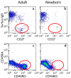
Naïve B- and T-lymphocytes in umbilical cord blood. In the upper panels, blood mononuclear cells were stained with CD19, IgD, and CD27 antibodies. B-lymphocytes were gated on CD19, and expression of surface IgD was plotted against CD27. In adult peripheral blood (panel (a)), memory B-lymphocytes, characterized by the phenotype surface IgD CD27+, are enclosed in the red circle. Memory B-lymphocytes in cord blood are virtually absent (panel (b)). In the lower panels, mononuclear cells were stained with CD3, CD45RA, and CD45RO. T-lymphocytes were gated on CD3, and memory T-lymphocytes, characterized by the phenotype CD45RA CD45RO+, are abundant in adult blood (panel (c)) and almost absent in cord blood (panel (d)). Data from GT Rijkers, unpublished.
Figure 3
Figure 3
Expression of CD21 on human marginal zone B cells. Immunohistochemistry of the HB5 (anti-CD21) antibody on sections of a spleen of a 12-month-old infant (panel (A)) and a 13-year-old child (panel (B)). In panel (B), the marginal zone (MZ) B cells show strong expression of CD21, but the infant MZ B cells (panel (A)) are lacking CD21 expression. GC, germinal center; C, corona; R, red pulp. Courtesy of Prof. W. Timens. The photograph in panel (B) has been published before [55].
Figure 4
Figure 4
Major developmental factors in microbial colonization. The progressive colonization of the infant gut is shown under the categorizations of natal status. From left to right, the figure depicts the progressive colonization of the gut with commensal (gray), probiotic (green), or pathogenic (red) bacteria. The progression of tight junction maturation is also presented (purple). Up arrow indicates increase of relative abundance, down arrow decrease of relative abundance.
Figure 5
Figure 5
Microbiota metabolites with an impact on the functionality of the immune system. Panel (a) show that various bacteria are able to produce the short-chain fatty acid butyrate as a byproduct of the fermentation of indigestible carbohydrates. Butyrate inhibits histone deacetylase (HDAC) which leads to the activation of FOXP3 in regulatory T cells. Butyrate also activates CD8+ cytotoxic T cells which produce IFN-γ and granzymes. Dietary tryptophan can be metabolized by L. reuteri into indole-3-aldehyde (I3A) (panel (b)) or by L. johnsonii and subsequently C. sporogenes into indole-3-propionic acid (IPA) (panel (c)). I3A, via unknown mechanisms, activates CD8+ cytotoxic T cells, while IPA leads to the activation of the Tcf7 transcription factor, resulting in the expansion of progenitor exhausted CD8+ cells (Tpex). See text for further explanation.
Figure 6
Figure 6
Immunoglobulin levels and gut microbiome development in early human life: IgG, IgM, and IgA levels are shown across fetal development, birth, and the first 1000 days postnatally. During the fetal period, IgG levels rise due to maternal transfer via the placenta. Postnatally, IgG levels initially decline as maternal antibodies are metabolized, but they gradually increase as the infant’s immune system matures and begins endogenous IgG production. IgM and IgA levels are present in smaller amounts, with both rising at a slower rate compared to IgG. The line representing bacterial diversity and abundance in the infant gut microbiome demonstrates a rapid postnatal expansion. This increase begins with early colonization and continues through the first 1000 days.

References

    1. Sansonetti P.J. War and peace at mucosal surfaces. Nat. Rev. Immunol. 2004;4:953–964. doi: 10.1038/nri1499. - DOI - PubMed
    1. Cupedo T., Crellin N.K., Papazian N., Rombouts E.J., Weijer K., Grogan J.L., Fibbe W.E., Cornelissen J.J., Spits H. Human fetal lymphoid tissue-inducer cells are interleukin 17-producing precursors to RORC+ CD127+ natural killer-like cells. Nat. Immunol. 2009;10:66–74. doi: 10.1038/ni.1668. - DOI - PubMed
    1. Ross C., Boroviak T.E. Origin and function of the yolk sac in primate embryogenesis. Nat. Commun. 2020;11:3760. doi: 10.1038/s41467-020-17575-w. - DOI - PMC - PubMed
    1. Enders A.C. Implantation in the macaque: Expansion of the implantation site during the first week of implantation. Placenta. 2007;28:794–802. doi: 10.1016/j.placenta.2006.11.001. - DOI - PubMed
    1. Xie X., Gou F., Zheng Z., Zhang Y., Zhang Y., Dong F., Cheng T., Cheng H. Decoding human bone marrow hematopoietic stem and progenitor cells from fetal to birth. iScience. 2024;27:110445. doi: 10.1016/j.isci.2024.110445. - DOI - PMC - PubMed

LinkOut - more resources