. 2025 Mar 12;14(6):416.
doi: 10.3390/cells14060416.

Differential Regulation of PKM2, AMPK, and mTOR in Response to Insulin and Dietary Management

Affiliations

Differential Regulation of PKM2, AMPK, and mTOR in Response to Insulin and Dietary Management

Emily Broberg et al. Cells. .

Abstract

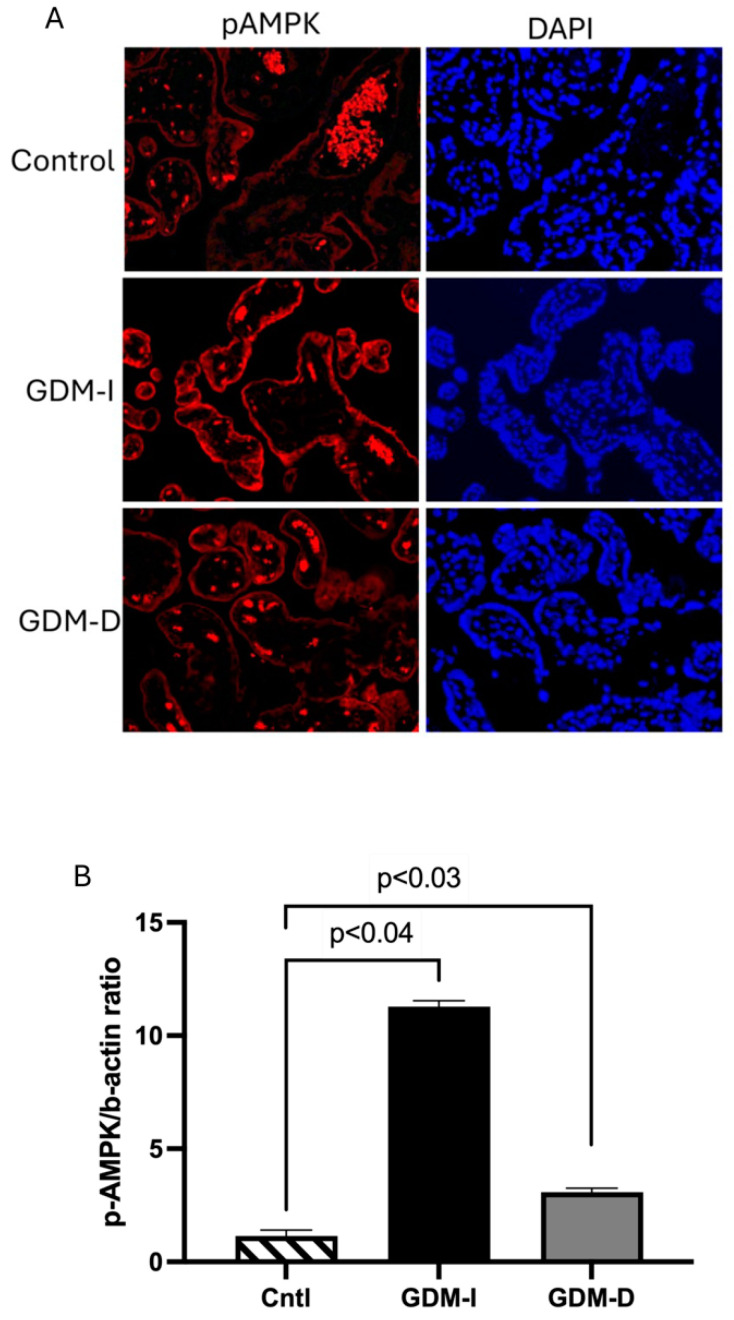
Gestational diabetes mellitus (GDM) affects placental metabolism, influencing both maternal and fetal outcomes. This study investigated the expression of metabolic regulators-Pyruvate Kinase M2 (PKM2), AMP-activated protein kinase (AMPK), and mTOR pathway components-in placental tissues from GDM pregnancies managed with either insulin (GDM-I) or dietary interventions (GDM-D). We hypothesize that metabolic adaptation in GDM is differentially regulated by treatment modality. This study analyzed 30 cases, including 10 control pregnancies,10 GDM-D cases, and 10 GDM-I cases. Analytical methods included immunofluorescence and immunoblotting. We observed an upregulation of PKM2 in both GDM-I and GDM-D placentas, suggesting enhanced glycolytic adaptation under GDM-induced metabolic stress. AMPK expression was significantly elevated in GDM-I and moderately increased in GDM-D placentas, potentially compensating for insulin resistance by promoting glucose uptake and energy homeostasis. Furthermore, mTOR pathway activation differed by treatment type, suggesting a treatment-specific mTOR response. The metabolic changes observed suggest that treatment modality in GDM may have direct implications for maternal and fetal health. Our findings indicate that while insulin and dietary management support metabolic adaptation in GDM, they do so through distinct mechanisms. These findings support a personalized approach in GDM treatment, where patient-specific metabolic responses should guide therapeutic decisions.

Keywords: AMPK; GDM; PKM2; mTOR; placental metabolism.


Conflict of interest statement

The authors declare they have no financial or non-financial conflicts of interest.

Figures

Figure 1
Figure 1
Placental mTOR pathway during GDM-I and GDM-D. Placental pmTOR (A), pp70 (B), and p4EBP1staining were performed between the control and disease placenta. Increased p-mTOR expression was only observed in the GDM-I placenta (A). pp70 was decreased in the GDM-D with no differences for the GDM-I (B). p4EBP1 was increased in GDM-D and decreased in GDM-I placentas compared to controls (C). Slides were viewed on a BX61 fluorescent microscope (20×) using the appropriate excitation and emission filters (fluorescein or rhodamine filters).
Figure 2
Figure 2
PKM2 expression and localization in the GDM-I and GDM-D placentas. The GDM-I placentas showed a higher syncytiotrophoblast expression of PKM2 than the GDM-D placentas. Slides were viewed on a BX61 fluorescent microscope (20×) using the appropriate excitation and emission filters with scale bars set at 100 μm.
Figure 3
Figure 3
Characteristic western blot of proteins studied.
Figure 4
Figure 4
PKM2, pPKM2, and pERK protein in the GDM-I and GDM-D placentas. Immunoblot showed an increase in PKM2 in GDM-I and GDM-D compared to controls (A,B). A higher PKM2 fold change was observed in the GDM-I placentas compared to the GDM-D tissues (C). Placental pPKM2 was decreased in the GDM-I and GDM-D tissues compared to controls (D,E). The decrease in the GDM-D placental tissues was more pronounced than in the GDM-I tissue (F). pERK was decreased in the GDM-I placentas (G), while no differences were observed for pERK in the GDM-D placentas (H). GDM-D pERK expression is higher than in the GDM-I placental tissues (I). Data are shown with p ≤ 0.05 when compared to controls.
Figure 5
Figure 5
Activated (p) placental AMPK in GDM-I and GDM-D patients. Immunoblot was performed for pAMPK protein determination. pAMPK protein was activated in the syncytiotrophoblast of the GDM-I placentas, and no differences were observed in the GDM-D placentas (A,B). Data are shown with significance of p ≤ 0.05 when compared to controls. Slides were viewed on a BX61 fluorescent microscope (20×) using the appropriate excitation and emission filters (fluorescein or rhodamine filters).

References

    1. Aires M.B., Dos Santos A.C. Effects of maternal diabetes on trophoblast cells. World J. Diabetes. 2015;6:338–344. doi: 10.4239/wjd.v6.i2.338. - DOI - PMC - PubMed
    1. Magee T.R., Ross M.G., Wedekind L., Desai M., Kjos S., Belkacemi L. Gestational diabetes mellitus alters apoptotic and inflammatory gene expression of trophobasts from human term placenta. J. Diabetes Complicat. 2014;28:448–459. doi: 10.1016/j.jdiacomp.2014.03.010. - DOI - PMC - PubMed
    1. Mejia C., Lewis J., Jordan C., Mejia J., Ogden C., Monson T., Winden D., Watson M., Reynolds P.R., Arroyo J.A. Decreased activation of placental mTOR family members is associated with the induction of intrauterine growth restriction by secondhand smoke in the mouse. Cell Tissue Res. 2017;367:387–395. doi: 10.1007/s00441-016-2496-5. - DOI - PubMed
    1. Plows J.F., Stanley J.L., Baker P.N., Reynolds C.M., Vickers M.H. The Pathophysiology of Gestational Diabetes Mellitus. Int. J. Mol. Sci. 2018;19:3342. doi: 10.3390/ijms19113342. - DOI - PMC - PubMed
    1. Modzelewski R., Stefanowicz-Rutkowska M.M., Matuszewski W., Bandurska-Stankiewicz E.M. Gestational Diabetes Mellitus-Recent Literature Review. J. Clin. Med. 2022;11:5736. doi: 10.3390/jcm11195736. - DOI - PMC - PubMed

Publication types

LinkOut - more resources