. 2025 Mar 20;14(6):461.
doi: 10.3390/cells14060461.

Efficacy of Deferoxamine Mesylate in Serum and Serum-Free Media: Adult Ventral Root Schwann Cell Survival Following Hydrogen Peroxide-Induced Cell Death

Affiliations

Efficacy of Deferoxamine Mesylate in Serum and Serum-Free Media: Adult Ventral Root Schwann Cell Survival Following Hydrogen Peroxide-Induced Cell Death

Yee Hang Ethan Ma et al. Cells. .

Abstract

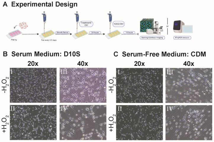
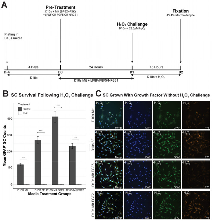
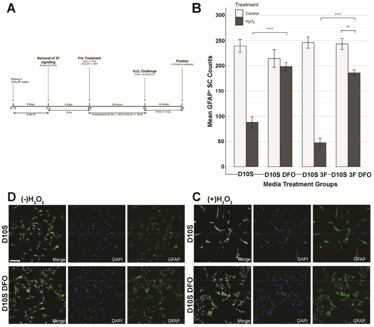
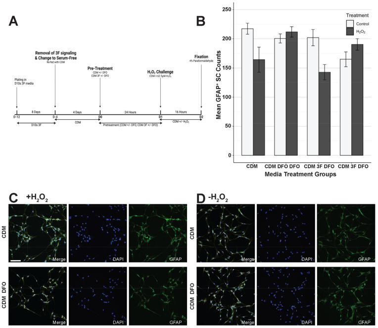
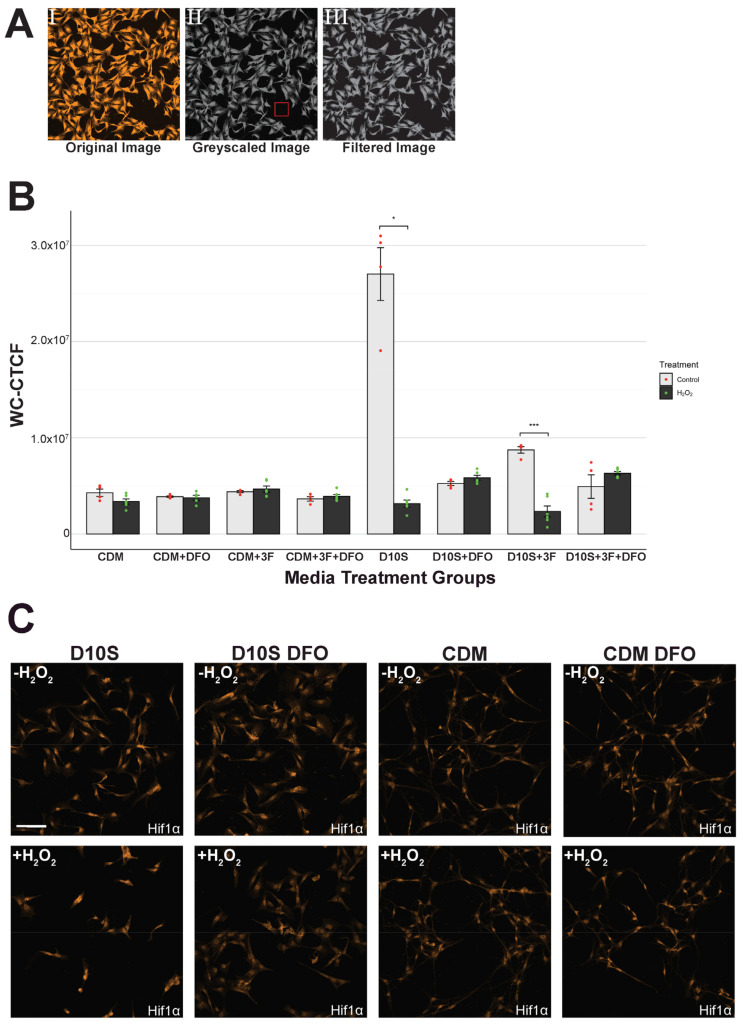
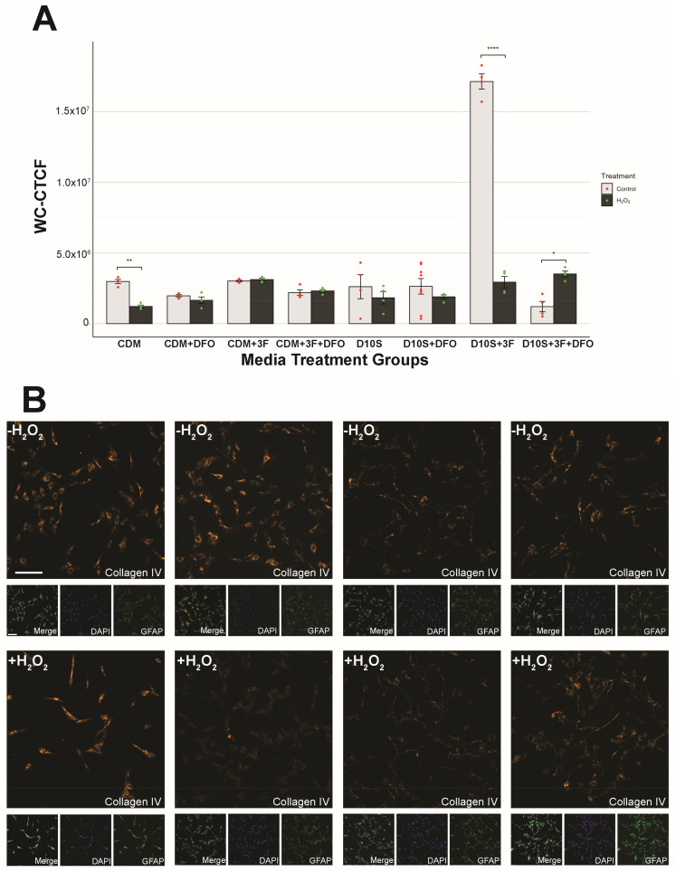
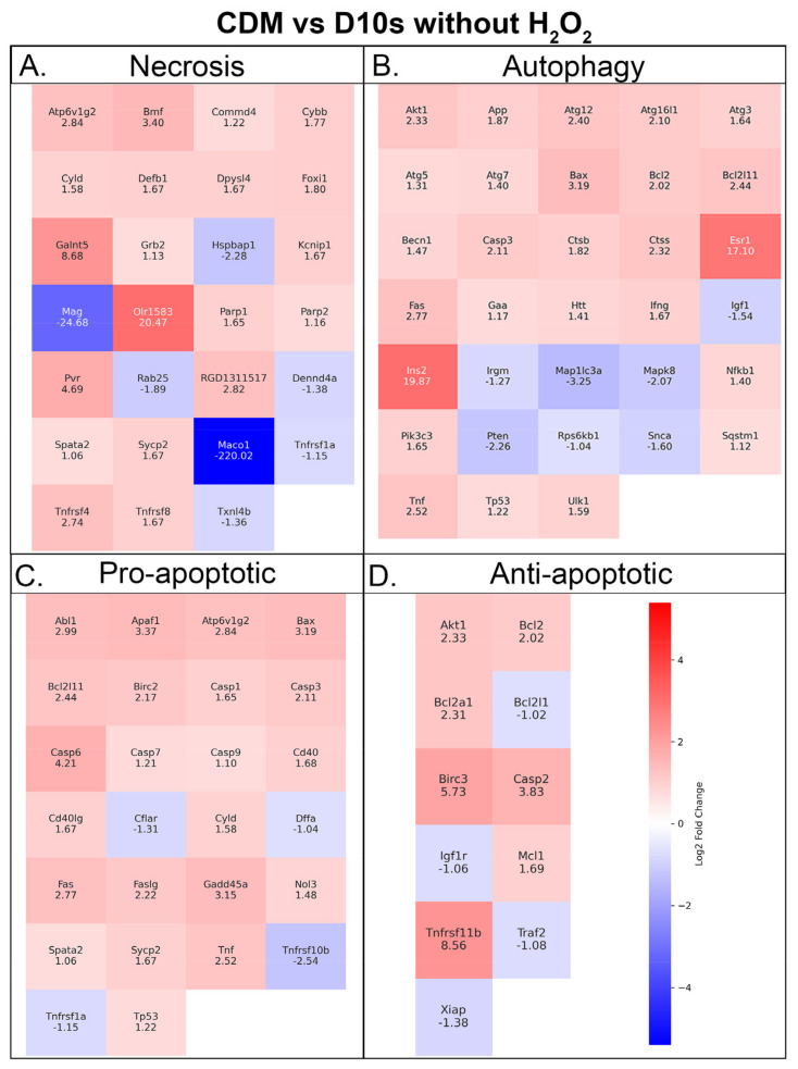
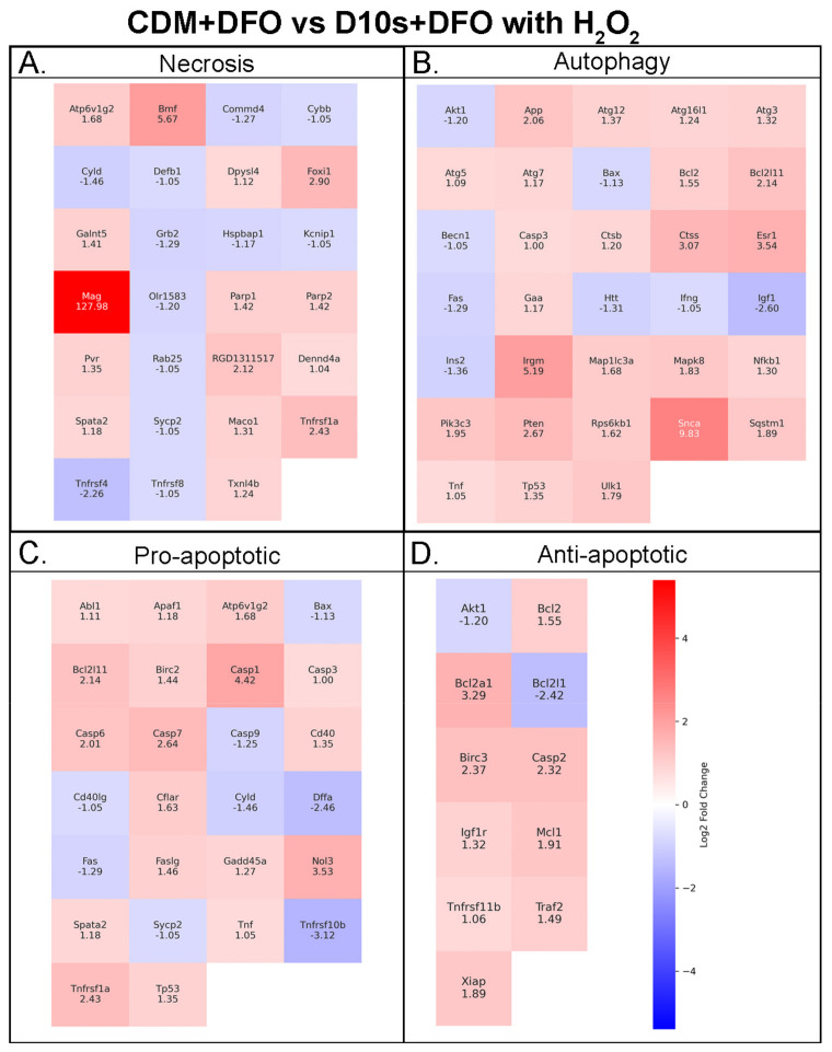
Schwann cell (SC) transplantation shows promise in treating spinal cord injury as a pro-regenerative agent to allow host endogenous neurons to bridge over the lesion. However, SC transplants face significant oxidative stress facilitated by ROS in the lesion, leading to poor survival. deferoxamine mesylate (DFO) is a neuroprotective agent shown to reduce H2O2-induced cell death in serum-containing conditions. Here we show that DFO is not necessary to induce neuroprotection under serum-free conditions by cell survival quantification and phenotypic analysis via immunohistochemistry, Hif1α and collagen IV quantification via whole cell corrected total cell fluorescence, and cell death transcript changes via RT-qPCR. Our results indicate survival of SC regardless of DFO pretreatment in serum-free conditions and an increased survival facilitated by DFO in serum-containing conditions. Furthermore, our results showed strong nuclear expression of Hif1α in serum-free conditions regardless of DFO pre-treatment and a nuclear expression of Hif1α in DFO-treated SCs in serum conditions. Transcriptomic analysis reveals upregulation of autophagy transcripts in SCs grown in serum-free media relative to SCs in serum conditions, with and without DFO and H2O2. Thus, indicating a pro-repair and regenerative state of the SCs in serum-free conditions. Overall, results indicate the protectiveness of CDM in enhancing SC survival against ROS-induced cell death in vitro.

Keywords: Schwann cells; deferoxamine mesylate; oxidative stress; spinal cord injury; transplantation.


Conflict of interest statement

The authors declare no conflicts of interest.

Figures

Figure 1
Figure 1
Schematic of experimental design and Schwann cell cultures. (A) Schematic of the experiments. Timepoints of treatments and hydrogen peroxide challenge. (B) Representative brightfield images of Schwann cells 16 h following hydrogen peroxide challenge (B(III,IV)) or sham (B(I,II)) in serum-containing medium. (B(I,III)) shows a 20× image, and (B(II,IV)) shows a 40× image. (C) Representative brightfield images of Schwann cells 16 h following hydrogen peroxide challenge (C(III,IV)) or sham (C(I,II)) in serum-free medium. (C(I,III)) shows a 20× image and (C(II,IV)) shows a 40× image. All images are taken on the Invitrogen EVOS XL CORE inverted brightfield microscope using EAchro 40× LWD PH, 0.65 NA/3.1 WD (AMEP4635), and Achro 20× LWD PH, 0.40 NA/6.8 WD (AMEP4934) objectives.
Figure 2
Figure 2
Growth factors do not protect against H2O2-induced Schwann cell death in vitro. (A) Schematic of the experiment. SCs were grown for 4 days on four-well chambers prior to exposure to treatment conditions. (B) Quantification of cellular survival in sham versus hydrogen peroxide challenged SCs in D10S Mit, D10S 3F, and D10S Mit + FGF2, D10S Mit + FGF5 shows no effect of growth factors in increasing SC survival. (C) Representative images of sham SCs in different conditions. Images are taken at x40. Channels: DAPI (405, blue), GFAP (488, green), P75 (Cy3, orange). Scale bar = 50 µm. * (p ≤ 0.05), ** (p ≤ 0.01), *** (p ≤ 0.001), **** (p ≤ 0.0001).
Figure 3
Figure 3
DFO increases Schwann cell survival in serum-containing medium. (A) Schematic of the experimental design. SCs were grown for 8 days in D10S 3F medium to eliminate fibroblasts in culture and promote SC survival and proliferation. (B) Quantification of GFAP + SC nucleus 16 h following sham or hydrogen peroxide challenge. D10S + DFO survival is significantly higher compared to D10S. D10S 3F + DFO survival is significantly higher than D10S 3F. (C) Representative images of D10S and D10S + DFO following 16 h of 62.5 μM hydrogen peroxide challenge. Channels: DAPI (405/blue), GFAP (488, green), Hif1α (cy3, orange). (D) Representative images of D10S and D10S + DFO following 16 h of sham. Channels: DAPI (405/blue), GFAP (488, green), Hif1α (cy3, orange). Hif1α staining is shown in Figure 5. Images taken at 20× magnification and 1.5× zoom. Scale bar = 100 μm. * (p ≤ 0.05), ** (p ≤ 0.01), *** (p ≤ 0.001), **** (p ≤ 0.0001).
Figure 4
Figure 4
Serum-free medium protects Schwann cells in H2O2-induced cell death. (A) Schematic of the experiment. SCs were grown for 8 days in D10S 3F medium to eliminate fibroblasts in culture and promote SC survival and proliferation. (B) Quantification of GFAP + SC nucleus 16 h following sham or hydrogen peroxide challenge. CDM + DFO survival shows no significant increase compared to CDM. CDM 3F + DFO survival shows no significant difference compared to CDM 3F. (C) Representative images of CDM and CDM + DFO following 16 h of 62.5 μM hydrogen peroxide challenge. Channels: DAPI (405/blue), GFAP (488, green), Hif1α (cy3, orange). (D) Representative images of CDM 3F and CDM 3F + DFO following 16 h of sham. Channels: DAPI (405, blue), GFAP (488, green), Hif1α (cy3, orange). Hif1α staining is shown in Figure 5. Images taken at 20× magnification and 1.5× zoom. Scale = 100 μm. * (p ≤ 0.05), ** (p ≤ 0.01), *** (p ≤ 0.001), **** (p ≤ 0.0001).
Figure 5
Figure 5
Hif1α WC-CTCF reveals serum variability. (A) Representative process of fluorescence intensity quantification. (A(I)) Original image without processing. (A(II)) Greyscale image, box shows definition of “background”. (A(III)) Image after background filtering to eliminate noise. (B) Quantification of Hif1α expression across groups. Statistics show high variability in serum groups with significant upregulation of Hif1α in non-DFO pretreatment groups. (C) Representative images of Hif1α expression across groups. Images taken at 20× magnification and 1.5× zoom. Scale = 100 μm. * (p ≤ 0.05), ** (p ≤ 0.01), *** (p ≤ 0.001), **** (p ≤ 0.0001).
Figure 6
Figure 6
Collagen IV WC-CTCF shows Schwann cell states. (A) Quantification of collagen IV expression. p values for statistics are shown in Supplemental Table S7. (B) Representative images of collagen IV expression across groups following 16 h of 62.5 μM hydrogen peroxide challenge or sham. Channels: Collagen IV (Cy3, orange), GFAP (488, green), DAPI (405, blue). Scale = 100 μm. * (p ≤ 0.05), ** (p ≤ 0.01), *** (p ≤ 0.001), **** (p ≤ 0.0001).
Figure 7
Figure 7
RT-qPCR shows increased metabolism and autophagy in CDM medium. Heatmaps showing Schwann cells in serum-free versus serum medium without the presence of H2O2, and transcript expression associated with (A) necrosis, (B) autophagy, (C) pro-apoptotic, and (D) antiapoptotic pathways. Fold-change values are shown on the heatmap.
Figure 8
Figure 8
RT-qPCR shows pro-repair and pro-regenerative Schwann cells in CDM media in the presence of DFO and H2O2. Heatmaps showing Schwann cells in serum-free versus serum medium with the presence of DFO and H2O2, and transcript expression associated with (A) necrosis, (B) autophagy, (C) pro-apoptotic, and (D) antiapoptotic pathways. Fold-change values are shown on the heatmap.

Similar articles

Cited by

References

    1. Bennett J., Das J.M., Emmady P.D. StatPearls [Internet] StatPearls Publishing; Treasure Island, FL, USA: 2024. [(accessed on 23 August 2024)]. Spinal Cord Injuries. Available online: https://www.ncbi.nlm.nih.gov/books/NBK560721/
    1. O’Shea T.M., Burda J.E., Sofroniew M.V. Cell biology of spinal cord injury and repair. J. Clin. Investig. 2017;127:3259–3270. doi: 10.1172/JCI90608. - DOI - PMC - PubMed
    1. Tran A.P., Warren P.M., Silver J. The Biology of Regeneration Failure and Success After Spinal Cord Injury. Physiol. Rev. 2018;98:881–917. doi: 10.1152/physrev.00017.2017. - DOI - PMC - PubMed
    1. Hill C.E. A view from the ending: Axonal dieback and regeneration following SCI. Neurosci. Lett. 2017;652:11–24. doi: 10.1016/j.neulet.2016.11.002. - DOI - PubMed
    1. Bunge M.B., Pearse D.D. Transplantation strategies to promote repair of the injured spinal cord. J. Rehabil. Res. Dev. 2003;40((Suppl. 1)):55–62. doi: 10.1682/JRRD.2003.08.0055. - DOI - PubMed

Publication types

MeSH terms

Substances

LinkOut - more resources