

The Effect of MRI Exposure on the Shear Bond Strength and Adhesive Remnant Index of Different Bracket Types
- PMID: 40136736
- PMCID: PMC11941266
- DOI: 10.3390/dj13030108
The Effect of MRI Exposure on the Shear Bond Strength and Adhesive Remnant Index of Different Bracket Types
Abstract
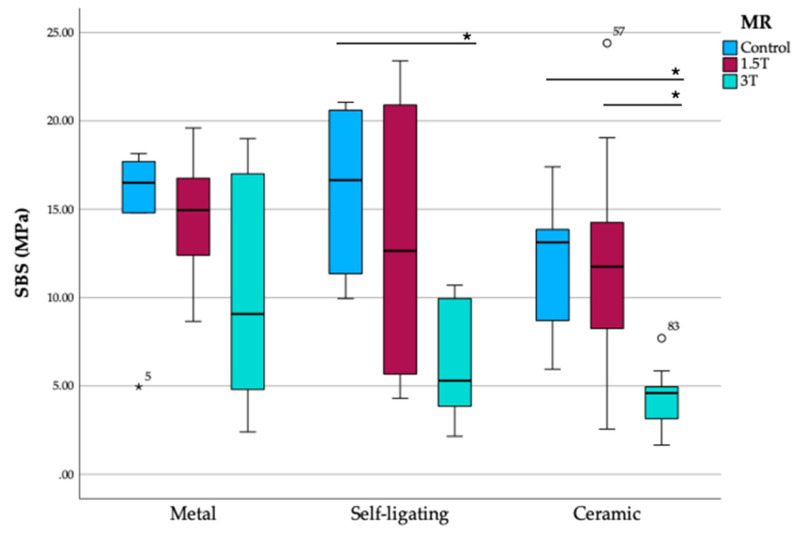
Background/Objectives: Magnetic resonance imaging (MRI) is widely used in diagnostics, but its effects on orthodontic materials remain a concern. This study aimed to evaluate the impact of MRI exposure at 1.5 T and 3 T on the shear bond strength (SBS) and adhesive remnant index (ARI) of different orthodontic bracket types (metal, self-ligating, and ceramic). Methods: A total of 90 extracted human premolars were divided into three groups (control, 1.5 T, and 3 T MRI exposure). The three bracket types were bonded using Transbond XT adhesive and subjected to standardized polymerization. MRI scans were conducted using 1.5 T and 3 T machines with clinically relevant sequences. SBS was measured using a universal testing machine, and the ARI was assessed under a stereomicroscope. Statistical analysis was performed using Kruskal-Wallis and chi-square tests. Results: MRI exposure influenced SBS and the ARI differently across bracket types. Firstly, 3 T MRI exposure significantly reduced SBS in self-ligating (p = 0.017) and ceramic brackets (p = 0.014) compared to the control, whereas metal brackets showed no significant changes. ARI scores varied across MRI conditions, with metal and self-ligating brackets showing increased adhesive retention at higher field strengths. No significant differences were observed in ARI scores for ceramic brackets across MRI conditions. Conclusions: The clinical importance of understanding these results is that both patients and clinicians must be aware of inevitable changes that occur in SBS during MRI, since exposure to high-field MRI, particularly 3 T, may alter bond strength and adhesive failure characteristics.
Keywords: adhesive remnant index; magnetic resonance imaging; orthodontic brackets; shear bond strength.
Conflict of interest statement
The authors declare no conflict of interest.
Figures
References
LinkOut - more resources
Full Text Sources
