. 2025 Mar 9;11(3):211.
doi: 10.3390/jof11030211.

Intra-Leaf Variability of Incubation Period Sheds New Light on the Lifestyle of Cercospora beticola in Sugar Beets

Affiliations

Intra-Leaf Variability of Incubation Period Sheds New Light on the Lifestyle of Cercospora beticola in Sugar Beets

Erich-Christian Oerke et al. J Fungi (Basel). .

Abstract

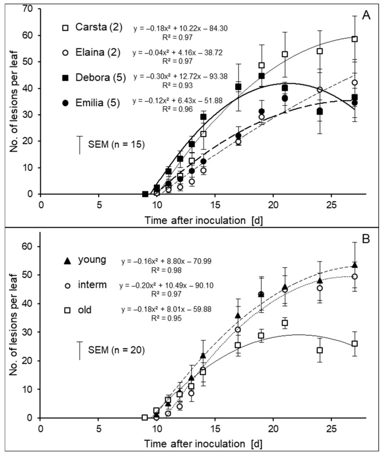
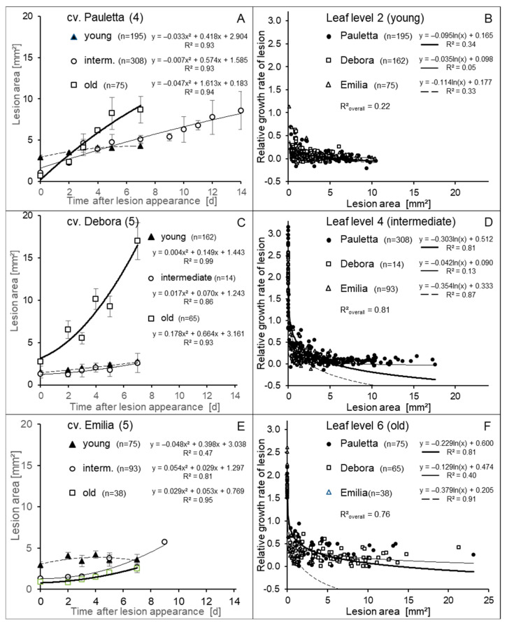
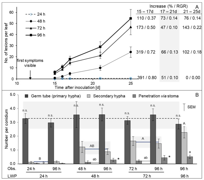
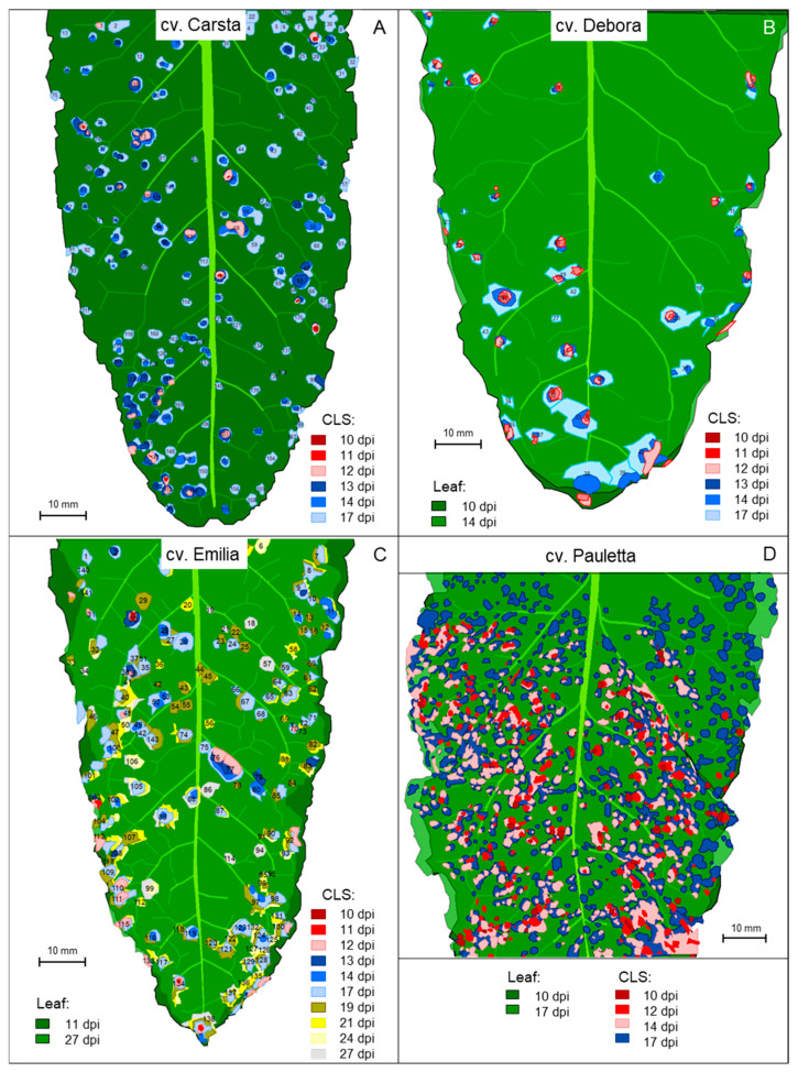
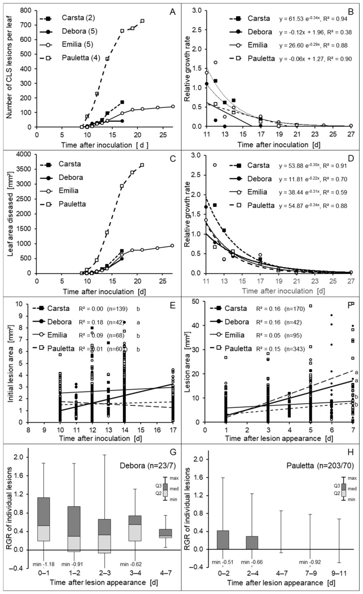
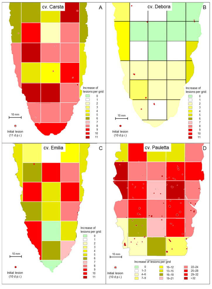
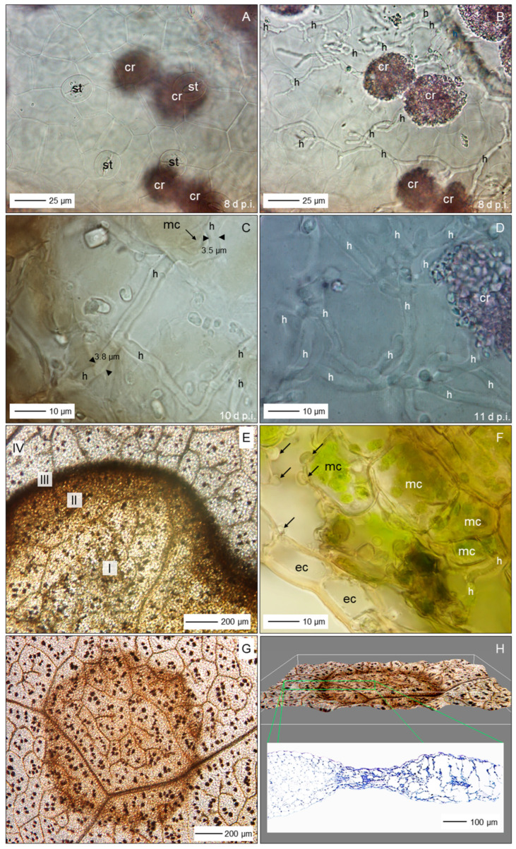
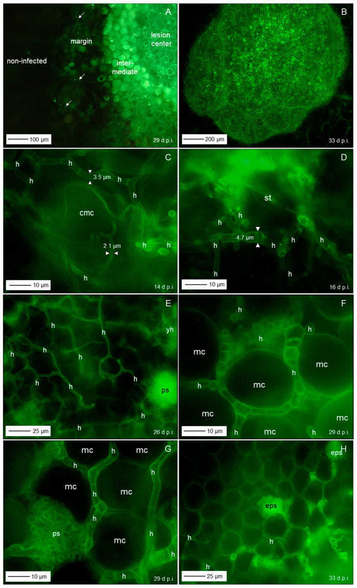
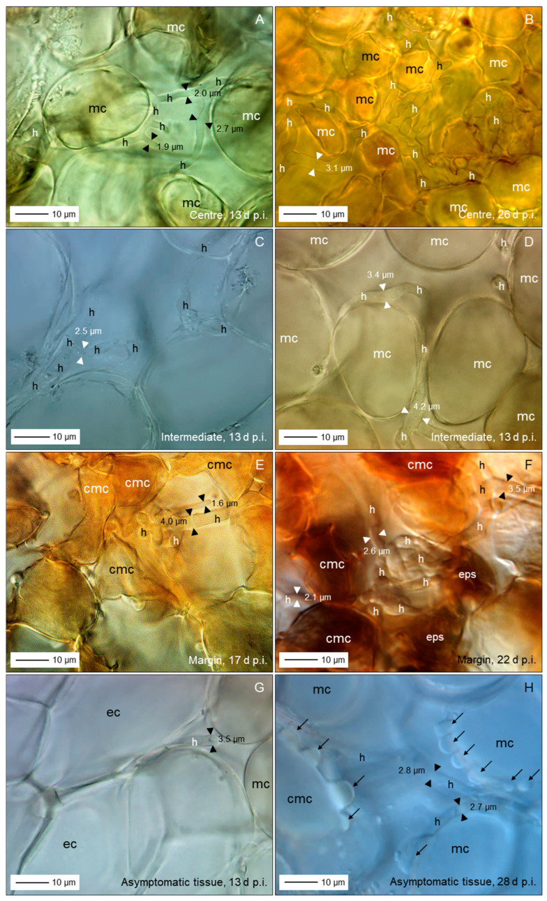
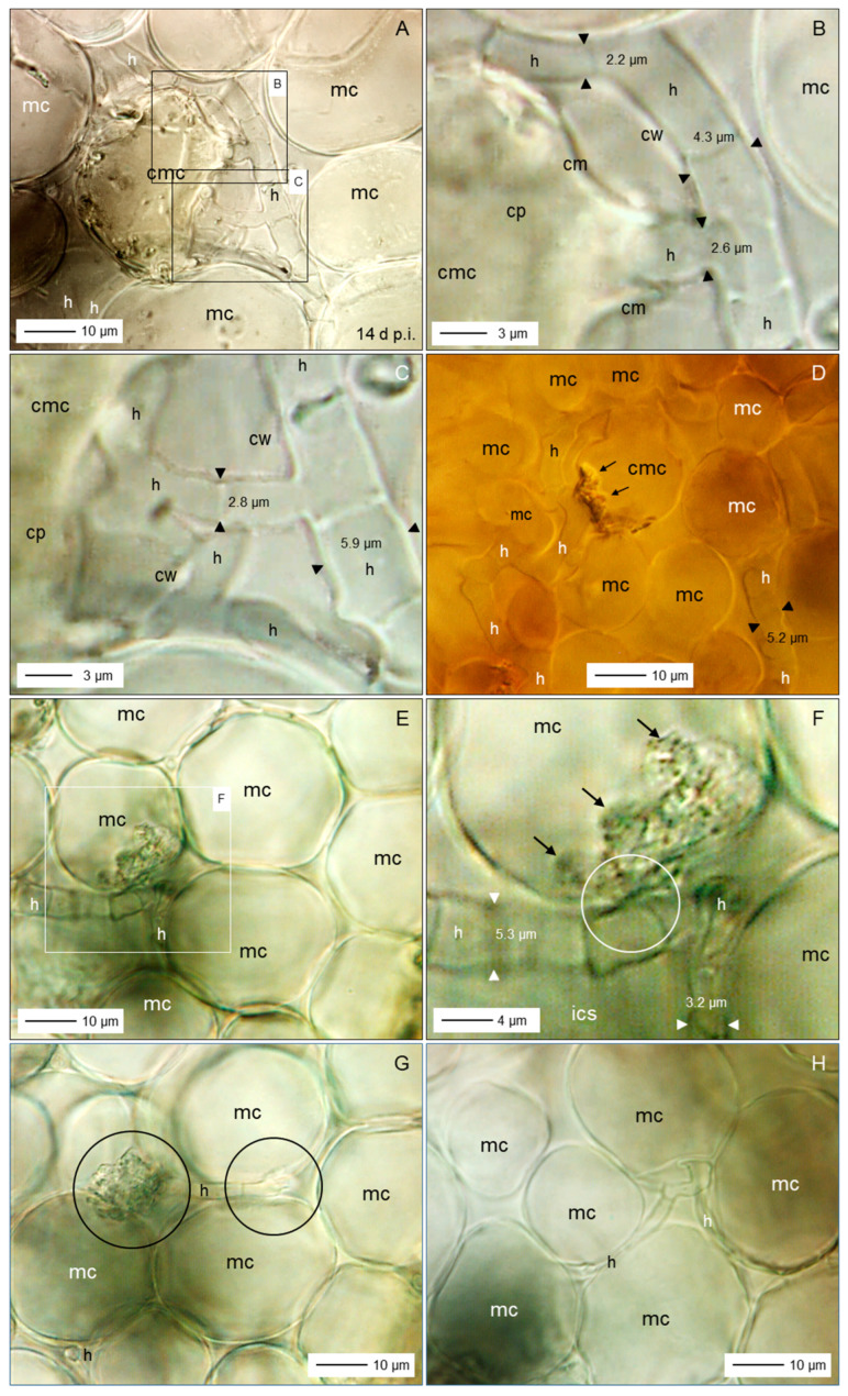
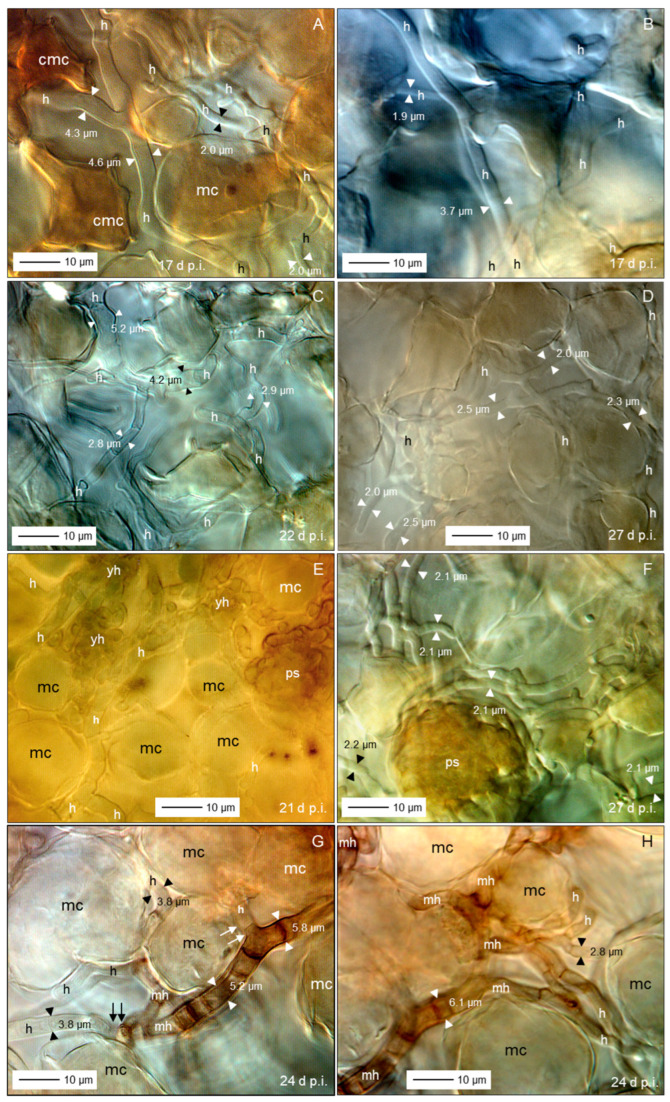
The length of incubation period, i.e., the time between first contact of host and pathogen and the appearance of symptoms, varies among diseases and depends on environmental conditions. Cercospora beticola is the most important fungal pathogen in sugar beet production worldwide, as Cercospora leaf spot (CLS) reduces the leaf area contributing to yield formation. Using sugar beet cultivars differing in CLS resistance, a single infection period of C. beticola resulted in minor differences in the incubation period among host genotypes and among individual plants of cultivars, greater differences among leaves within plants, and substantial variation within individual leaves. Under greenhouse conditions not suitable for secondary infections, the first CLS lesions appeared 10 days after inoculation; however, the number of leaf spots and CLS severity further increased significantly for another 7 to 17 days. A geographic information system approach enabled the tracking of colony appearance and growth of all CLSs on inoculated leaves for up to 27 days. Asymptomatic colonization of leaves was associated with thick hyphae which switched to thin hyphae or melanization after lesion appearance. The lifestyle of C. beticola-intercellular tissue colonization, triggering of necrotic host reaction-is discussed considering the experimental results as well as literature resources.

Keywords: asymptomatic colonization; hemibiotrophy; hyphal dimorphism; necrotrophy; toxin.


Conflict of interest statement

The authors declare no conflicts of interest.

Figures

Figure 1
Figure 1
Increase in the number of CLS lesions of sugar beet leaves depending on the disease susceptibility of cultivars (A) and the ontogenetic stage of leaves (B). Two cultivars with low (score 2) and moderate (score 5) CLS susceptibility, respectively, and three leaf ages (young; level 2 from top); intermediate, level 4 from trop; old, level 6 from top) were assessed for typical CLS lesions over a period of 8 to 27 d p.i. (4 cvs. × 3 leaf levels × 5 replicates).
Figure 2
Figure 2
Growth of CLS lesions after the time of first appearance depending on sugar beet cultivar and leaf age (A,C,E) and relationship between lesion size and relative growth rate (RGR; for two days) of lesions depending on leaf age (B,D,F).
Figure 3
Figure 3
Effect of length of initial leaf wetness period on the number of CLS lesions 15 to 25 d p.i. and increase in the number of lesions over time expressed as % increase and relative growth rate (RGR), respectively (cv. Brix, n = 33; (A)). Growth of C. beticola on the leaf surface; formation of primary and secondary germ tubes and successful penetration via stomata 24, 48, 72, and 96 h p.i. (B). Grey background visualizes the range of parameter variability (n = 30; Obs., Time of leaf sampling; LWP, leaf wetness period). Different letters represent statistical significant (p ≤0.05) differences between average values for 24, 48, 72, and 96 h leaf wetness duration (upper case letters for secondary hyphae, lowercase letters for penetration via stoma); asterisk indicates significant difference (p ≤ 0.05) between observations made directly after leaf wetness and 96 h, respectively.
Figure 4
Figure 4
Appearance and development of individual CLS lesions on leaves of four sugar beet cultivars; (A) cv. Carsta in the period 10 to 17 d p.i.; (B) cv. Debora in the period 10 to 17 d p.i.; (C) cv. Emilia in the period 10 to 27 d p.i.; (D) cv. Pauletta in the period 10 to 17 d p.i. The colour code represents the time of lesion appearance; the size of lesions represents lesion growth in time. As shape and area of leaves were affected by disease and time, two leaf outlines are given for the leaf of cvs. Debora, Emilia, and Pauletta, respectively. Numbers within lesions indicate the numbering of lesions during data processing.
Figure 5
Figure 5
Development of CLS lesions on leaves of four sugar beet cultivars in time. Increase in the number of CLS lesions per leaf in the period 9 to 27 d p.i. (A); relative growth rate (RGR) of lesion number over time (B); increase in the total leaf area diseased in the period 9 to 27 d p.i. (C); relative growth rate (RGR) of diseased leaf area (D); effect of the length of incubation period on the initial area of CLS lesions (E); growth of CLS lesions within the period of 7 days after the time of appearance (F); box-and-whisker plots of the relative growth rate of individual CLS lesions on cv. Debora (G) and Pauletta (H) in response to the time after lesion appearance. Assessment of lesions appearing 10 and 11 d p.i. (cv. Debora) and 10 and 12 d p.i. (cv. Pauletta), respectively. Lower case letters indicate significant differences (p ≤ 0.05) among cultivars (E,F).
Figure 6
Figure 6
Spatial patterns of CLS on individual leaves of four sugar beet varieties; (A) cv. Carsta; (B) cv. Debora; (C) cv. Emilia; (D) cv. Pauletta. First lesions (red dots) 10 d p.i. (cv. Carsta 11 d p.i.) and spatial variability in the increase of CLS lesions within the period 10 to 17 d p.i. Colour coding represents the intensity of formation of additional CLS lesions per grid within a period of 7 days.
Figure 7
Figure 7
Characteristics of sugar beet leaves infected by C. beticola. Surface of cleared leaf tissue without disease symptoms, 8 d p.i.; st, stoma; cr, crystal inclusions (A); same site with focus a little bit lower demonstrating intensive colonization by C. beticola hyphae (h; (B)); close-up of non-melanized intercellular hyphae with a diameter about double that of epicuticular germ tubes (C,D); subareas of sugar beet leaf tissue with a CLS lesion; I, lesion centre; II, intermediate; III, (brown) margin; IV, green leaf area outside the lesion (E); cross section of sugar beet tissue at the margin of a CLS lesion 14 d p.i.; epidermal cells (ec) and mesophyll cells (mc; with chloroplasts) form globular protuberances (F); top view (G) and side view (H) of a CLS lesion visualizing tissue breakdown of the margin and the less pronounced damage of the lesion centre. Bright-field microscopy after staining with toluidine blue ((AD,H), lower part).
Figure 8
Figure 8
Colonization of leaf tissue by C. beticola. Subareas of CLS lesion and neighbouring tissue; arrows point to hyphal structure of margin and beyond (A); total lesion with cellular organization of leaf tissue, 33 d p.i. (B); hyphae with diameter ranging from 3 to 5 µm intercellularly colonized the lesion centre (C,D,F) eventually forming early pseudostromata above the mesophyll tissue (E,G,H). Epifluorescence microscopy after staining with aniline blue. cmc, collapsed mesophyll cell; eps, early pseudostroma; h, hypha; mc, mesophyll cell; st, stoma; yh, yeast-like hypha.
Figure 9
Figure 9
Differences in the tissue colonization of subareas of CLS lesions early after CLS lesion appearance (A,C,D,E,G) and in a later stage of pathogenesis (B,F,H). Intensity of intercellular colonization of leaf centre increased with time (A,B); intercellular hyphae in the intermediate region hardly damaged mesophyll cells (C,D); intercellular growth of hyphae among necrotic cells of the reddish-brown margin increased with time (E,F); leaf tissue outside the CLS lesion had minimal intercellular hyphae and hardly any cell damage (G,H). Bright-field microscopy after tissue clearing with chloral hydrate. cmc, collapsed mesophyll cell; ec, epidermal cell; eps, early pseudostroma; h, hypha; mc, mesophyll cell.
Figure 10
Figure 10
Details on intercellular growth of C. beticola hyphae in the centre of CLS lesions, 14 (AD) and 17 (EH) d p.i. Hyphae (h) with a diameter >5 µm grew between mesophyll cells that reacted to cell penetration by (thinner) hyphae with collapse of individual cells; neighbouring cells were not affected. cmc, collapsed mesophyll cell; cp, cytoplasm; cw, cell wall; mc, ics, intercellular space; mesophyll cell. Squares indicate to areas presented at higher magnification in the next image(s); circles and arrows highlight interesting areas and structures, respectively.
Figure 11
Figure 11
Variability of C. beticola hyphae growing in the centre of CLS lesions. In early lesions, hyphae with a diameter > 3.5 µm predominated (A,B). Hyphae formed after lesion appearance were narrower and became predominant in older lesions (C,D). The formation of early pseudostromata was associated with the formation of yeast-like shorter hyphae (yh), which became melanized in later stages of leaf colonization (E,F). Filamentous intercellular hyphae became melanized as well; they formed thinner, non-melanized hyphae which penetrated mesophyll cells (G,H). Bright-field microscopy after tissue clearing with chloral hydrate. Arrows indicate the transition from melanized to non-melanized hyphal cells.

Similar articles

References

    1. Shane W., Teng P. Impact of Cercospora leaf spot on root weight, sugar yield, and purity of Beta vulgaris. Plant Dis. 1992;76:812–820. doi: 10.1094/PD-76-0812. - DOI
    1. Groenewald M., Groenewald J.Z., Harrington T., Abeln E.C.A., Crous P.W. Mating type gene analysis in apparently asexual Cercospora species is suggestive of cryptic sex. Fungal Genet. Biol. 2006;43:813–825. doi: 10.1016/j.fgb.2006.05.008. - DOI - PubMed
    1. Groenewald J., Nakashima C., Nishikawa J., Shin H.D., Park J.H., Jama A., Groenewald M., Braun U., Crous P.W. Species concepts in Cercospora: Spotting the weeds among the roses. Stud. Mycol. 2013;75:115–170. doi: 10.3114/sim0012. - DOI - PMC - PubMed
    1. Rangel L.I., Spanner R.E., Ebert M.K., Pethybridge S.J., Stukenbrock E.H., de Jonge R., Secor G.A., Bolton M.D. Cercospora beticola: The intoxicating lifestyle of the leaf spot pathogen of sugar beet. Mol. Plant Pathol. 2020;21:1020–1041. doi: 10.1111/mpp.12962. - DOI - PMC - PubMed
    1. Weiland J., Koch G. Sugarbeet leaf spot disease (Cercospora beticola Sacc.) Mol. Plant Pathol. 2004;5:157–166. doi: 10.1111/j.1364-3703.2004.00218.x. - DOI - PubMed

LinkOut - more resources