Review
. 2025 Mar 14;16(3):105.
doi: 10.3390/jfb16030105.

Three-Dimensional Bioprinting for Intervertebral Disc Regeneration

Affiliations
Review

Three-Dimensional Bioprinting for Intervertebral Disc Regeneration

Md Amit Hasan Tanvir et al. J Funct Biomater. .

Abstract

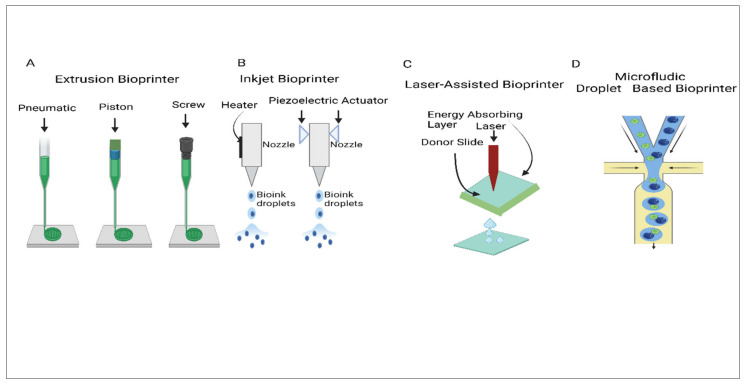
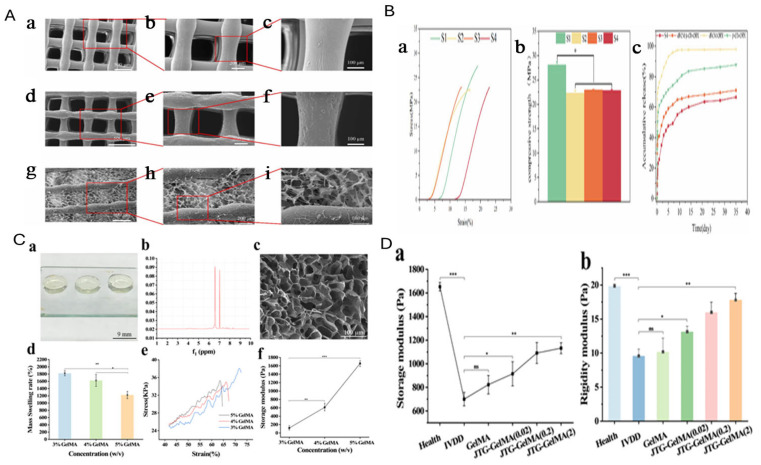
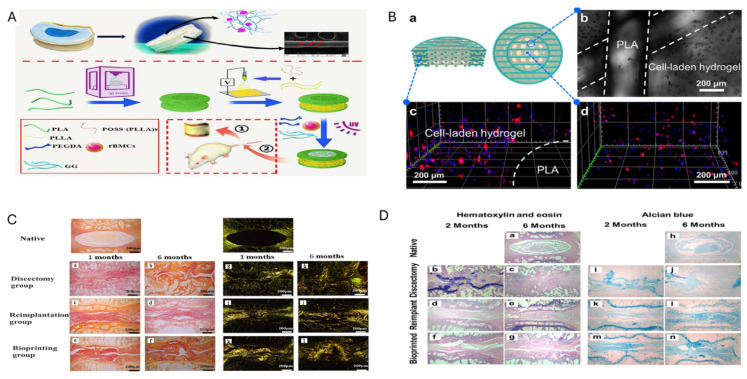
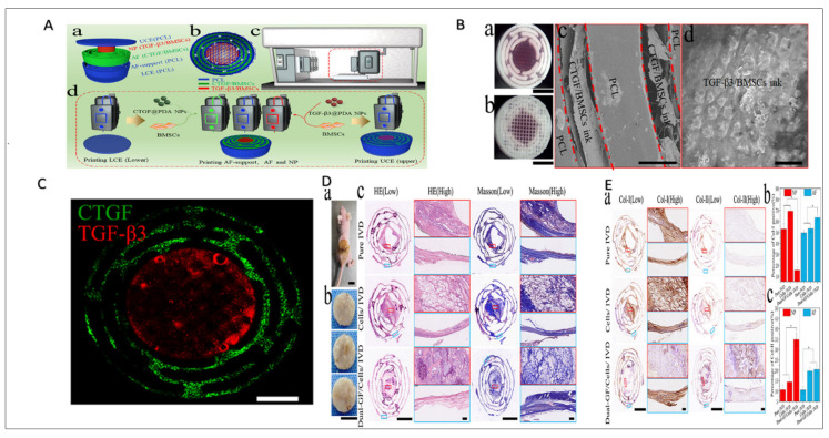
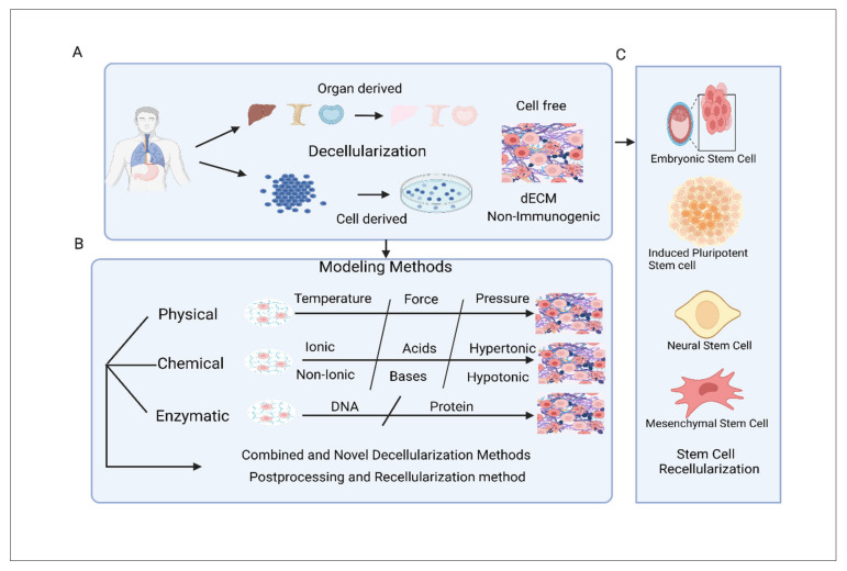
The rising demand for organ transplants and the need for precise tissue models have positioned the in vitro biomanufacturing of tissues and organs as a pivotal area in regenerative treatment. Considerable development has been achieved in growing tissue-engineered intervertebral disc (IVD) scaffolds, designed to meet stringent mechanical and biological compatibility criteria. Among the cutting-edge approaches, 3D bioprinting stands out due to its unparalleled capacity to organize biomaterials, bioactive molecules, and living cells with high precision. Despite these advancements, polymer-based scaffolds still encounter limitations in replicating the extracellular matrix (ECM)-like environment, which is fundamental for optimal cellular activities. To overcome these challenges, integrating polymers with hydrogels has been recommended as a promising solution. This combination enables the advancement of porous scaffolds that nurture cell adhesion, proliferation, as well as differentiation. Additionally, bioinks derived from the decellularized extracellular matrix (dECM) have exhibited potential in replicating biologically relevant microenvironments, enhancing cell viability, differentiation, and motility. Hydrogels, whether derived from natural sources involving collagen and alginate or synthesized chemically, are highly valued for their ECM-like properties and superior biocompatibility. This review will explore recent advancements in techniques and technologies for IVD regeneration. Emphasis will be placed on identifying research gaps and proposing strategies to bridge them, with the goal of accelerating the translation of IVDs into clinical applications.

Keywords: 3D bioprinting; IVD; biomaterials; dECM.


Conflict of interest statement

The authors declare no conflicts of interest.

Figures

Figure 8
Figure 8
(A) SEM images of freeze-dried GelMA hydrogel at three different concentrations: (a) 5% GelMA, showing a porous structure with small, interconnected pores; (b) 10% GelMA, displaying a denser network with reduced pore size; (c) 15% GelMA, revealing a compact structure with minimal porosity. (B) Immunofluorescence staining of NPCs in GelMA hydrogels: (A1–a) Immunofluorescence staining of NPCs 7 days after encapsulation at 5% concentrations GelMA hydrogels; (A1–b) Immunofluorescence staining of NPCs 7 days after encapsulation at 10% concentrations GelMA hydrogels; (A1–c) Immunofluorescence staining of NPCs 7 days after encapsulation at 15% concentrations GelMA hydrogels; (B1) Quantitative optical density analysis of Col II expression. (** p < 0.01). Reproduced and adapted with permission from [177]. (C) Immunofluorescence staining for Col I and TNMD expression in TGF-β1-treated groups: (a) Col I/DAPI staining at days 7 and 14 in PCL, PCL/3% GelMA, and PCL/3% GelMA+TGFβ1 groups, showing increased Col I expression in response to TGFβ1; (b) TNMD/DAPI staining at days 7 and 14, indicating enhanced TNMD expression under TGFβ1 treatment. Reproduced and adapted with permission from [178]. (D) Morphology and ECM expression of NPMSCs were assessed via aggrecan (a,b) and col-II (c,d) immunofluorescence staining and quantification across three hydrogel types. Reproduced and adapted with permission from [183].
Figure 1
Figure 1
Overview of the 3D bioprinting workflow for regenerative medicine: (i) identification of defect via medical imaging, (ii) customized implant design using computer-aided tools, (iii) expansion of autologous stem cells in culture, (iv) formulation of bio-ink with biomaterials and cells, (v) fabrication of the 3D construct via bioprinting, (vi) cell maturation under specific conditions, and (vii) implantation of the matured construct into the defect site.
Figure 2
Figure 2
Timeline highlighting major milestones in 3D bioprinting advancements, from the invention of SLA in 1984 to the bioprinting of functional human tissues and organs by 2019.
Figure 3
Figure 3
The bioprinting method for creating 3D tissues predictably starts with picturing the injured tissue and environment to direct the design of the printed tissue. Design methodologies such as biomimicry, tissue self-assembly, and MT building blocks are possible to apply individually. Picking the right materials and cell sources is crucial, tailored to the specific form and function of the target tissue. Commonly used materials comprise synthetic or natural polymers and decellularized ECM. These components must work with bioprinting techniques, for example, inkjet, microextrusion, or laser-assisted printing. Certain bioprinted tissues may demand a maturation phase in a bioreactor before they are ready for transplantation, while others can be applied directly for in vitro purposes. Reproduction and adapted with permission from [12] copyright 2014 Nature.
Figure 4
Figure 4
Different schematic diagrams of 3D bioprinting.
Figure 5
Figure 5
(A) SEM images of different scaffold structures at various magnifications, showing S1 (a–c). S2 (d–f). S4 (g–i). (a,d and g) 100×, (b, e and h) 200×, and (c, f and i) 500×, morphology and porosity. Reproduced and adapted with permission from ref [129] under copyright 2012 RSC Advance. (B) Mechanical characterization of scaffolds: (a) stress–strain curves, (b) compressive modulus comparison, and (c) time-dependent mechanical behaviour. Reproduced and adapted with permission from ref [129] copyright 2012 RSC Advance. (C) Material characterization: (a) macroscopic view, (b) FTIR analysis, (c) porous morphology under SEM, and (d–f) water absorption, swelling behaviour, and degradation profile. Reproduced and adapted with permission from ref [130] copyright 2010 Elsevier. (D) Biological evaluation: (a) storage modulus comparison and (b) relative cell viability of different scaffold formulations. Reproduced and adapted with permission from ref [130]. Copyright 2010 Elsevier. (level of significance is * p < 0.05, ** p < 0.01, and *** p < 0.001) (ns: No significance).
Figure 6
Figure 6
(A) A diagrammatic representation of the method involved in initiating a biomimetic artificial IVD) scaffold applying a mixture of 3D printing and electrospinning practices. Reproduced and adapted with permission from [155] copyright 2005 Elsevier. (B) Laser Scanning Confocal Microscopy images of the constructs after 14 days of culture: (a) Schematic of the bioprinted IVD construct, showing structural and cell-laden components; (b) High-magnification image displaying the PLA structure and cell-laden hydrogel interface; (c) Confocal image of the cell-laden hydrogel, showing cell distribution and viability; (d) 3D reconstruction of cell-laden hydrogel and PLA scaffold, illustrating cellular organization. Reproduced and adapted with permission from [154] copyright 2012 Elsevier. (C) Sirius red staining and Polarized Light Microscopy of disc sections. Native disc tissue at 1 and 6 months, showing intact collagen fibers: (a,b,g,h) Discectomy group, revealing collagen degradation; (c,d,i,j) Reimplantation group, showing partial collagen restoration; (e,f,k,l) The Bioprinting group demonstrates improved collagen alignment. Reproduced and adapted with permission from [155]. (D) Hematoxylin & Eosin (H&E) and Alcian Blue Staining: (a) Native disc sections at 2 and 6 months, showing normal tissue structure; (b,c) Discectomy group, displaying tissue degeneration; (d,e) Reimplantation group, indicating partial structural recovery; (f,g) Bioprinting group, showing enhanced disc restoration alcian blue; (h) Native disc section; (i,j) Discectomy group, displaying tissue degeneration; (k,l) Reimplantation group, indicating slightly structural recovery; (m,n) Bioprinting group, demonstrating significant proteoglycan retention. Reproduced and adapted with permission from [154] copyright 2012 Elsevier.
Figure 7
Figure 7
The model was composed of UCE, LCE, NP, AF, and AF-support ((A), a), with UCE, LCE, and AF-support sections printed using PCL polymer, 3D bioprinter,and the print process of 3D-bioprinted IVD scaffold ((A), b–d). The dual growth factor (GF)–releasing IVD scaffold’s morphology and mechanical properties were analyzed, showing alignment with the designed structure ((B), a–d). Fluorescence pictures of TGF-β3 (red) and CTGF (green) in the 3D-bioprinted IVD scaffold (C). For in vivo reconstruction, pure IVD, cells/IVD, and dual-GFs/cells/IVD scaffolds were implanted in nude mice, revealing that cells/IVD and dual-GFs/cells/IVD scaffolds improved cartilage, collagen, and chondrocyte formation after 3 months ((D), a–c). Notably, the dual-GFs/cells/IVD scaffold in the NP region had higher Col II and lower Col I levels, while the AF region exhibited increased Col I and Col II compared to other scaffolds ((E), a–c). Reproduced and adapted with permission from ref [173] copyright 2020 Elsevier.
Figure 9
Figure 9
(A) Classification of dECM. (B) Different fabrication methods for creating dECM from cells and organs. (C) Recellularizing dECM scaffolds to create bioengineered grafts for tissue engineering.

Similar articles

References

    1. Melrose J. Strategies in regenerative medicine for intervertebral disc repair using mesenchymal stem cells and bioscaffolds. Regen. Med. 2016;11:705–724. doi: 10.2217/rme-2016-0069. - DOI - PubMed
    1. Khaleque M.A., Kim J.-H., Hwang B.-J., Kang J.-K., Quan M., Kim Y.-Y. Role of necroptosis in intervertebral disc degeneration. Int. J. Mol. Sci. 2023;24:15292. doi: 10.3390/ijms242015292. - DOI - PMC - PubMed
    1. Seo J.-Y., Kim J., Kim Y.-Y., Ha K.-Y., Kim Y.-H., Kim S.-I., Lim J.-H., Seo K.B., Kang H., Choi S. Autophagy in an extruded disc compared to the remaining disc after lumbar disc herniation in the same patient. Eur. Spine J. 2024;33:61–67. doi: 10.1007/s00586-023-07731-3. - DOI - PubMed
    1. Stolworthy D.K., Fullwood R.A., Merrell T.M., Bridgewater L.C., Bowden A.E. Biomechanical analysis of the camelid cervical intervertebral disc. J. Orthop. Transl. 2015;3:34–43. - PMC - PubMed
    1. Khaleque M.A., Kim J.-H., Lee H.-H., Kim G.-H., You W.-Y., Lee W.-J., Kim Y.-Y. Comparative Analysis of Autophagy and Apoptosis in Disc Degeneration: Understanding the Dynamics of Temporary-Compression-Induced Early Autophagy and Sustained-Compression-Triggered Apoptosis. Int. J. Mol. Sci. 2024;25:2352. doi: 10.3390/ijms25042352. - DOI - PMC - PubMed

LinkOut - more resources