

Calcified Lung Nodules: A Diagnostic Challenge in Clinical Daily Practice
- PMID: 40137568
- PMCID: PMC11946818
- DOI: 10.3390/tomography11030028
Calcified Lung Nodules: A Diagnostic Challenge in Clinical Daily Practice
Abstract
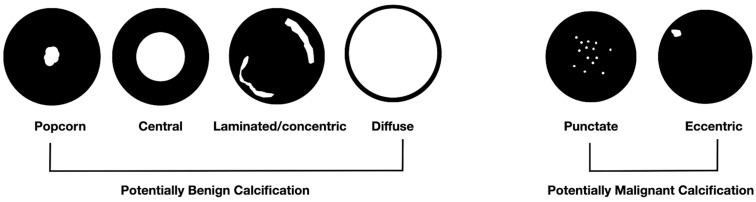
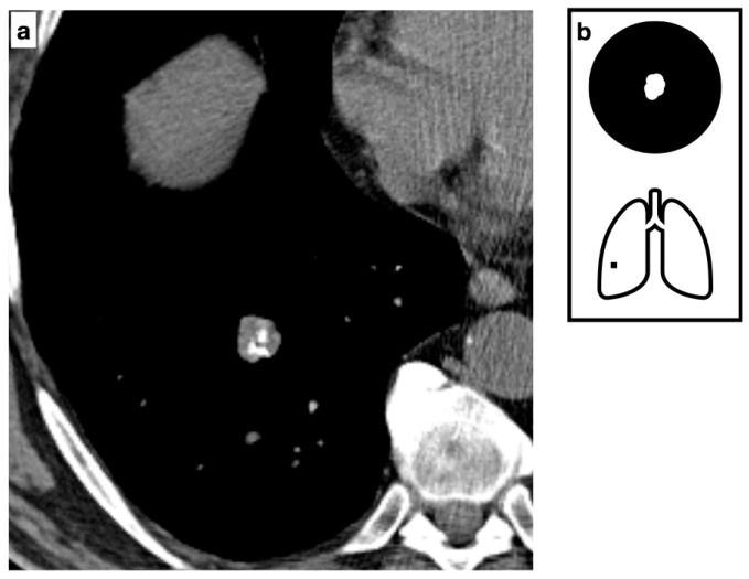
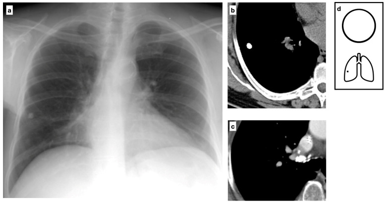
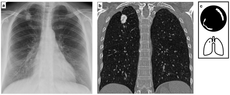
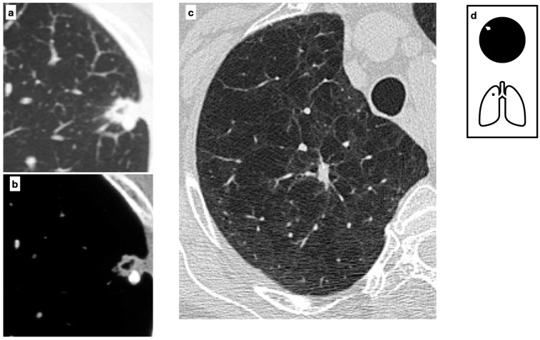
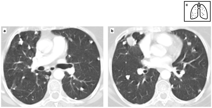
Calcified lung nodules are frequently encountered on chest imaging, often as incidental findings. While calcifications are typically associated with benign conditions, they do not inherently exclude malignancy, making accurate differentiation essential. The primary diagnostic challenge lies in distinguishing benign from malignant nodules based solely on imaging features. Various calcification patterns, including diffuse, popcorn, lamellated and eccentric, provide important diagnostic clues, though overlap among different conditions may persist. A comprehensive diagnostic approach integrates clinical history with multimodal imaging, including magnetic resonance and nuclear medicine, when necessary, to improve accuracy. When imaging findings remain inconclusive, tissue sampling through biopsy may be required for definitive characterization. This review provides an overview of the imaging features of calcified lung nodules, emphasizing key diagnostic challenges and their clinical implications.
Keywords: calcification pattern; chest CT; lung nodules; pulmonary calcifications.
Conflict of interest statement
The authors declare no conflicts of interest.
Figures
References
-
- Fiorelli A., D’andrilli A., Carlucci A., Vicidomini G., Argento G., Marinucci B.T., Ardissone F., Rapanà R., Sobrero S., Carbognani P., et al. Pulmonary Hamartoma Associated With Lung Cancer (PHALC Study): Results of a Multicenter Study. Lung. 2021;199:369–378. doi: 10.1007/s00408-021-00460-8. - DOI - PMC - PubMed
Publication types
MeSH terms
LinkOut - more resources
Full Text Sources
Medical
