

Diagnostic Sensitivity of the Revised Venous System in Brain Death in Children
- PMID: 40137570
- PMCID: PMC11945848
- DOI: 10.3390/tomography11030030
Diagnostic Sensitivity of the Revised Venous System in Brain Death in Children
Abstract
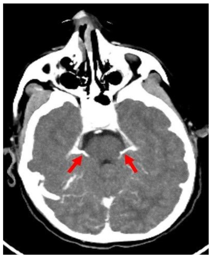
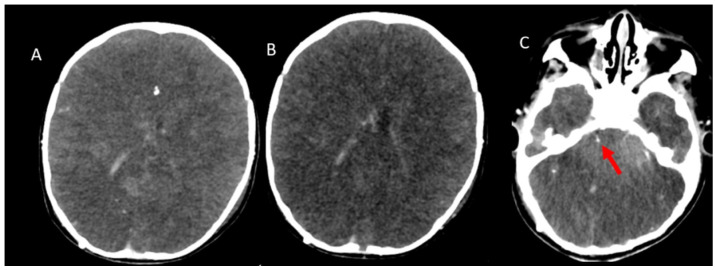
Background/objectives: While ancillary tests for brain death diagnosis are not routinely recommended in guidelines, they may be necessary in specific clinical scenarios. Computed tomography angiography (CTA) is particularly advantageous in pediatric patients due to its noninvasive nature, accessibility, and rapid provision of anatomical information. This study aims to assess the diagnostic sensitivity of a revised venous system (ICV-SPV) utilizing a 4-point scoring system in children clinically diagnosed with brain death.
Materials and methods: A total of 43 pediatric patients clinically diagnosed with brain death who underwent CTA were retrospectively analyzed. Imaging was performed using a standardized brain death protocol. Three distinct 4-point scoring systems (A20-V60, A60-V60, ICV-SPV) were utilized to assess vessel opacification in different imaging phases. To evaluate age-dependent sensitivity, patients were categorized into three age groups: 26 days-1 year, 2-6 years, and 6-18 years. The sensitivity of each 4-point scoring system in diagnosing brain death was calculated for all age groups.
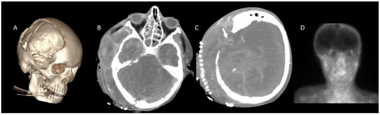
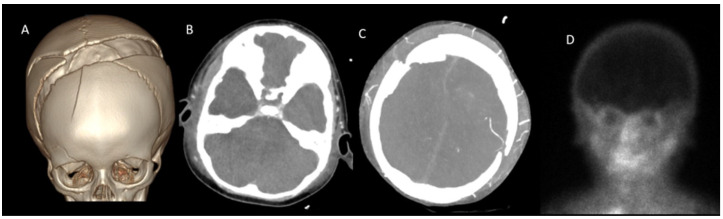
Results: The revised venous scoring system (ICV-SPV) demonstrated the highest overall sensitivity in confirming brain death across all age groups, significantly outperforming the reference 4-point scoring systems. Furthermore, the ICV-SPV system exhibited the greatest sensitivity in patients with cranial defects.
Conclusions: The revised 4-point venous CTA scoring system, which relies on the absence of ICV and SPV opacification, is a reliable tool for confirming cerebral circulatory arrest in pediatric patients with clinical brain death.
Keywords: angiography; brain death; children; computed tomography.
Conflict of interest statement
The authors declare no conflicts of interest.
Figures
Similar articles
-
Assessment of Cerebral Circulatory Arrest via CT Angiography and CT Perfusion in Brain Death Confirmation.Korean J Radiol. 2021 Mar;22(3):395-404. doi: 10.3348/kjr.2019.0859. Epub 2020 Sep 10. Korean J Radiol. 2021. PMID: 32932559 Free PMC article.
-
Revised CT angiography venous score with consideration of infratentorial circulation value for diagnosing brain death.Ann Intensive Care. 2016 Dec;6(1):88. doi: 10.1186/s13613-016-0188-7. Epub 2016 Sep 13. Ann Intensive Care. 2016. PMID: 27620878 Free PMC article.
-
Computed tomography angiography as a confirmatory test for the diagnosis of brain death.J Neurosurg. 2018 Feb;128(2):639-644. doi: 10.3171/2016.10.JNS161042. Epub 2017 Mar 17. J Neurosurg. 2018. PMID: 28304181
-
Ancillary testing in brain death.Semin Neurol. 2015 Apr;35(2):125-38. doi: 10.1055/s-0035-1547541. Epub 2015 Apr 3. Semin Neurol. 2015. PMID: 25839721 Review.
-
Computed Tomography Angiography (CTA) in Selected Scenarios with Risk of Possible False-Positive or False-Negative Conclusions in Diagnosing Brain Death.Life (Basel). 2022 Oct 6;12(10):1551. doi: 10.3390/life12101551. Life (Basel). 2022. PMID: 36294986 Free PMC article. Review.
References
-
- Greer D.M., Kirschen M.P., Lewis A., Gronseth G.S., Rae-Grant A., Ashwal S., Babu M.A., Bauer D.F., Billinghurst L., Corey A., et al. Pediatric and Adult Brain Death/Death by Neurologic Criteria Consensus Guideline. Neurology. 2023;101:1112–1132. doi: 10.1212/WNL.0000000000207740. - DOI - PMC - PubMed
-
- Nakagawa T.A., Ashwal S., Mathur M., Mysore M.R., Bruce D., Conway E.E., Jr., Duthie S.E., Hamrick S., Harrison R., Kline A.M., et al. Guidelines for the determination of brain death in infants and children: An update of the 1987 Task Force recommendations. Crit. Care Med. 2011;39:2139–2155. doi: 10.1097/CCM.0b013e31821f0d4f. - DOI - PubMed
-
- McKinnon N.K., Maratta C., Zuckier L.S., Boyd J.G., Chassé M., Hornby L., Kramer A., Kromm J., Mooney O.T., Muthusami P., et al. Ancillary investigations for death determination in infants and children: A systematic review and meta-analysis. Can. J. Anaesth. 2023;70:749–770. doi: 10.1007/s12630-023-02418-1. - DOI - PMC - PubMed
MeSH terms
LinkOut - more resources
Full Text Sources
