. 2025 Mar 14;11(3):35.
doi: 10.3390/tomography11030035.

Ground-Glass Opacities in the Access Route and Biopsy in Highly Perfused Dependent Areas of the Lungs as Risk Factors for Pulmonary Hemorrhage During CT-Guided Lung Biopsy: A Retrospective Study

Affiliations

Ground-Glass Opacities in the Access Route and Biopsy in Highly Perfused Dependent Areas of the Lungs as Risk Factors for Pulmonary Hemorrhage During CT-Guided Lung Biopsy: A Retrospective Study

Michael P Brönnimann et al. Tomography. .

Abstract

Background/objectives: The risk of hemorrhage during CT-guided lung biopsy has not been systematically studied in cases where ground-glass opacities (GGO) are present in the access route or when biopsies are performed in highly perfused, dependent lung areas. While patient positioning has been studied for pneumothorax prevention, its role in minimizing hemorrhage risk remains unexplored. This study aimed to determine whether GGOs in the access route and biopsies in dependent lung areas are risk factors for pulmonary hemorrhage during CT-guided lung biopsy.

Methods: A retrospective analysis was conducted on 115 CT-guided lung biopsies performed at a single center (2020-2023). Patients were categorized based on post-interventional hemorrhage exceeding 2 cm (Grade 2 or higher). We evaluated the presence of GGOs in the access route and biopsy location (dependent vs. non-dependent areas) using chi square, Fisher's exact, and Mann-Whitney U tests. Univariate and multivariate logistic regression analyses were conducted to evaluate risk factors for pulmonary hemorrhage.

Results: Pulmonary hemorrhage beyond 2 cm occurred in 30 of 115 patients (26%). GGOs in the access route were identified in 67% of these cases (p < 0.01), and hemorrhage occurred more frequently when biopsies were performed in dependent lung areas (63% vs. 40%, p = 0.03). Multivariable analysis showed that GGOs in the access route (OR 5.169, 95% CI 1.889-14.144, p = 0.001) and biopsies in dependent areas (OR 4.064, 95% CI 1.477-11.186, p < 0.001) independently increased hemorrhage risk.

Conclusions: GGOs in the access route and dependent lung area biopsies are independent risk factors for hemorrhage during CT-guided lung biopsy.

Keywords: biopsy; hemorrhage; image-guided biopsy; radiography interventional; risk factors; tomography.


Conflict of interest statement

The authors declare no conflicts of interest.

Figures
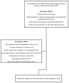
Figure 1
Figure 1
Flowchart showing the study population.
Figure 2
Figure 2
Technical implementation. (A) Lesion in the right upper lobe and ipsilateral-dependent patient positioning to prevent a pneumothorax. (B) After full core lung biopsy with adjustable penetration depth, a lobar hemorrhage develops (red-colored area).
Figure 3
Figure 3
A schematic representation of the zoning approach used in this study based on the position-dependent influence of gravity on pleural pressure (PPL). Pleural pressure increases progressively from the non-dependent upper lung regions to the dependent lower regions. Additionally, gravitational forces contribute to enhanced perfusion in the dependent lung areas [18,19,29]. To simplify classification, only the “RED” zone was designated as dependent. The zoning method followed the rule of thirds.
Figure 4
Figure 4
Ground-glass opacification (GGO) in the access route. (A) Asymmetric GGO in the access route peripheral to the target lesion. (BE) A classic subsolid nodule with surrounding GGO and Grade 2 hemorrhage after biopsy.
Figure 5
Figure 5
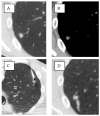
Lesion in the lower lobe without GGO in the access route. (A) Small 13 mm suspicious solid lesion in the right lower lobe. (B) Target lesion in the corresponding soft tissue window. (C) Prone positioning with biopsy. (D) Pulmonary hemorrhage Grade 1 in the access route.
Figure 6
Figure 6
Pulmonary hemorrhage grading system on CT scans performed before/during (left) and directly after (right) the biopsy pulmonary hemorrhage. (A) Grade 0: without image morphological evidence of parenchymal hemorrhage after biopsy. (B) Grade 1: new focal hemorrhage less than 2 cm beyond the needle path can be delineated in the access route after the biopsy. (C) Grade 2: after the biopsy, the target lesion can no longer be demarcated if the alveolar hemorrhage is locally larger than 2 cm. (D) Grade 3: lobar hemorrhage after biopsy. (E) Grade 4: hemothorax with immediate hyperdense fluid within the pleural cavity after biopsy [23].
Figure 7
Figure 7
Histological findings of the lung biopsies. HVL = hypervascular lesions; other HVM = hypervascular metastasis like sarcoma; SCLC = small cell lung cancer; NSCLC = non-small cell lung cancer; HCC = hepatocellular carcinoma; NET = neuroendocrine tumor; RCC = renal cell carcinoma. Percentages are rounded. Metastases from breast cancer and RCC are not included in the pie chart as no cases were recorded.
Figure 8
Figure 8
Histological findings of lung biopsies with GGO in the access route. Total number = 46; 40% of all performed lung biopsies. The numbers in the pie chart correspond to the frequency. GGO = ground-glass opacity; other HVM = hypervascular metastasis like sarcoma; SCLC = small cell lung cancer; NSCLC = non-small cell lung cancer; HCC = hepatocellular carcinoma; NET = neuroendocrine tumor; RCC = renal cell carcinoma. No cases were recorded for NSCLC, HCC, RCC, or breast cancer metastases.

Similar articles

References

    1. Nour-Eldin N.-E.A., Alsubhi M., Naguib N.N., Lehnert T., Emam A., Beeres M., Bodelle B., Koitka K., Vogl T.J., Jacobi V. Risk factor analysis of pulmonary hemorrhage complicating CT-guided lung biopsy in coaxial and non-coaxial core biopsy techniques in 650 patients. Eur. J. Radiol. 2014;83:1945–1952. - PubMed
    1. Tai R., Dunne R.M., Trotman-Dickenson B., Jacobson F.L., Madan R., Kumamaru K.K., Hunsaker A.R. Frequency and severity of pulmonary hemorrhage in patients undergoing percutaneous CT-guided transthoracic lung biopsy: Single-institution experience of 1175 cases. Radiology. 2016;279:287–296. doi: 10.1148/radiol.2015150381. - DOI - PubMed
    1. Tomiyama N., Yasuhara Y., Nakajima Y., Adachi S., Arai Y., Kusumoto M., Eguchi K., Kuriyama K., Sakai F., Noguchi M. CT-guided needle biopsy of lung lesions: A survey of severe complication based on 9783 biopsies in Japan. Eur. J. Radiol. 2006;59:60–64. doi: 10.1016/j.ejrad.2006.02.001. - DOI - PubMed
    1. Heerink W.J., de Bock G.H., de Jonge G.J., Groen H.J., Vliegenthart R., Oudkerk M. Complication rates of CT-guided transthoracic lung biopsy: Meta-analysis. Eur. Radiol. 2017;27:138–148. - PMC - PubMed
    1. Yeow K.-M., Su I.-H., Pan K.-T., Tsay P.-K., Lui K.-W., Cheung Y.-C., Chou A.S.B. Risk factors of pneumothorax and bleeding: Multivariate analysis of 660 CT-guided coaxial cutting needle lung biopsies. Chest. 2004;126:748–754. doi: 10.1378/chest.126.3.748. - DOI - PubMed