

Discussion of a Simple Method to Generate Descriptive Images Using Predictive ResNet Model Weights and Feature Maps for Recurrent Cervix Cancer
- PMID: 40137578
- PMCID: PMC11946054
- DOI: 10.3390/tomography11030038
Discussion of a Simple Method to Generate Descriptive Images Using Predictive ResNet Model Weights and Feature Maps for Recurrent Cervix Cancer
Abstract
Background: Predictive models like Residual Neural Networks (ResNets) can use Magnetic Resonance Imaging (MRI) data to identify cervix tumors likely to recur after radiotherapy (RT) with high accuracy. However, there persists a lack of insight into model selections (explainability). In this study, we explored whether model features could be used to generate simulated images as a method of model explainability.
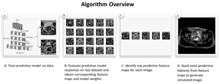
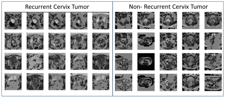
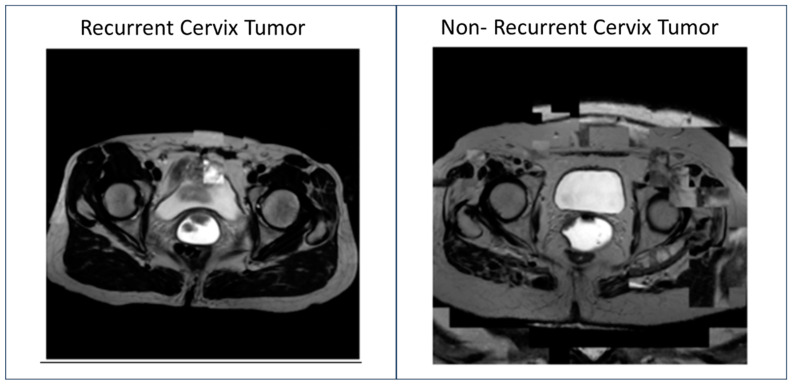
Methods: T2W MRI data were collected for twenty-seven women with cervix cancer who received RT from the TCGA-CESC database. Simulated images were generated as follows: [A] a ResNet model was trained to identify recurrent cervix cancer; [B] a model was evaluated on T2W MRI data for subjects to obtain corresponding feature maps; [C] most important feature maps were determined for each image; [D] feature maps were combined across all images to generate a simulated image; [E] the final image was reviewed by a radiation oncologist and an initial algorithm to identify the likelihood of recurrence.
Results: Predictive feature maps from the ResNet model (93% accuracy) were used to generate simulated images. Simulated images passed through the model were identified as recurrent and non-recurrent cervix tumors after radiotherapy. A radiation oncologist identified the simulated images as cervix tumors with characteristics of aggressive Cervical Cancer. These images also contained multiple MRI features not considered clinically relevant.
Conclusion: This simple method was able to generate simulated MRI data that mimicked recurrent and non-recurrent cervix cancer tumor images. These generated images could be useful for evaluating the explainability of predictive models and to assist radiologists with the identification of features likely to predict disease course.
Keywords: ResNet; XAI; cervix cancer; deep learning; generated images; machine learning; model explainability; most important feature maps; radiation therapy; radiotherapy.
Conflict of interest statement
The authors declare no conflicts of interest.
Figures
Similar articles
-
3cDe-Net: a cervical cancer cell detection network based on an improved backbone network and multiscale feature fusion.BMC Med Imaging. 2022 Jul 23;22(1):130. doi: 10.1186/s12880-022-00852-z. BMC Med Imaging. 2022. PMID: 35870877 Free PMC article.
-
Convolutional neural network to predict the local recurrence of giant cell tumor of bone after curettage based on pre-surgery magnetic resonance images.Eur Radiol. 2019 Oct;29(10):5441-5451. doi: 10.1007/s00330-019-06082-2. Epub 2019 Mar 11. Eur Radiol. 2019. PMID: 30859281
-
Predicting microvascular invasion in hepatocellular carcinoma: a deep learning model validated across hospitals.Cancer Imaging. 2021 Oct 9;21(1):56. doi: 10.1186/s40644-021-00425-3. Cancer Imaging. 2021. PMID: 34627393 Free PMC article.
-
Prediction of tumor control in patients with cervical cancer: analysis of combined volume and dynamic enhancement pattern by MR imaging.AJR Am J Roentgenol. 1998 Jan;170(1):177-82. doi: 10.2214/ajr.170.1.9423627. AJR Am J Roentgenol. 1998. PMID: 9423627
-
Recurrent uterine cervical carcinoma: spectrum of imaging findings.Korean J Radiol. 2000 Oct-Dec;1(4):198-207. doi: 10.3348/kjr.2000.1.4.198. Korean J Radiol. 2000. PMID: 11752955 Free PMC article. Review.
References
-
- Quinn M., Benedet J., Odicino F., Maisonneuve P., Beller U., Creasman W., Heintz A., Nan H., Pecorelli S. Carcinoma of the Cervix Uteri. Int. J. Gynecol. Obstet. 2006;95:S43–S103. - PubMed
-
- Chino J., Annunziata C.M., Beriwal S., Bradfield L., Erickson B.A., Fields E.C., Fitch K., Harkenrider M.M., Holschneider C.H., Kamrava M., et al. Radiation Therapy for Cervical Cancer: Executive Summary of an ASTRO Clinical Practice Guideline. Pract. Radiat. Oncol. 2020;10:220–234. doi: 10.1016/j.prro.2020.04.002. - DOI - PMC - PubMed
-
- Chen S., Chen Y., Yu L., Hu X. YTHDC1 inhibits cell proliferation and angiogenesis in cervical cancer by regulating m6 A modification of SOCS4 mRNA. Mol. Cell. Toxicol. 2024;20:533–540. doi: 10.1007/s13273-023-00360-3. - DOI
