. 2025 Mar 28;11(13):eadu8116.
doi: 10.1126/sciadv.adu8116. Epub 2025 Mar 26.

Histone modification-driven structural remodeling unleashes DNMT3B in DNA methylation

Affiliations

Histone modification-driven structural remodeling unleashes DNMT3B in DNA methylation

Chao-Cheng Cho et al. Sci Adv. .

Abstract

The DNA methyltransferase 3B (DNMT3B) plays a vital role in shaping DNA methylation patterns during mammalian development. DNMT3B is intricately regulated by histone H3 modifications, yet the dynamic interplay between DNMT3B and histone modifications remains enigmatic. Here, we demonstrate that the PWWP (proline-tryptophan-tryptophan-proline) domain within DNMT3B exhibits remarkable dynamics that enhances the enzyme's methyltransferase activity upon interactions with a modified histone H3 peptide (H3K4me0K36me3). In the presence of H3K4me0K36me3, both the PWWP and ADD (ATRX-DNMT3-DNMT3L) domains transition from autoinhibitory to active conformations. In this active state, the PWWP domain most often aligns closely with the catalytic domain, allowing for simultaneous interactions with H3 and DNA to stimulate DNA methylation. The prostate cancer-associated DNMT3B R545C mutant is even more dynamic and susceptible to adopting the active conformation, resulting in aberrant DNA hypermethylation. Our study suggests the mechanism by which conformational rearrangements in DNMT3B are triggered by histone modifications, ultimately unleashing its activity in DNA methylation.


Figures

Fig. 1.
Fig. 1.. The PWWP domain within DNMT3B is required for the histone H3 peptide–mediated stimulation of the methyltransferase activity of the DNMT3B-3L complex.
(A) Methyltransferase activities of 3BΔ214-3L in the absence or presence of the H3 tail peptide with methylated and/or unmethylated K4 and K36 were measured using MTase-Glo assays by three independent experiments. The 40–amino acid H3K4me0K36me3 peptide (residues 1 to 40) most strongly stimulated the methyltransferase activity of 3BΔ214-3L in methylating 40-bp DNA. MTase, methyltransferase. (B) The H3K4me0K36me3 peptide did not enhance the methyltransferase activity of 3BΔ412-3L, but it did increase the activity of 3BΔ214-3L. The average of three independent experiments is shown with error bars representing 1 SD. Statistical significance (P values) was determined by two-tailed Student’s t test: *P < 0.1 and ****P < 0.0001; ns, not significant.
Fig. 2.
Fig. 2.. The PWWP domain in the DNMT3B-3L complex is involved in DNA binding upon interactions with the H3K4me0K36me3 peptide.
(A) The DNA binding affinities of the 3BΔ214-3L complex in the absence and presence of the H3 tail peptide were measured by fluorescence polarization spectroscopy. (B) The DNA binding affinities of the PWWP domain and PWWP-K268A mutant in the absence and presence of the H3K4me0K36me3 peptide were measured by fluorescence polarization spectroscopy. Data are plotted as means ± SD from three independent experiments and fitted using GraphPad Prism version 9. (C) Superimposition of the crystal structures of apo-form PWWP (this study; PDB entry: 8ZLK) and H3-bound PWWP (PDB entry: 5CIU) reveals that the hydrogen bonding network between Asp266 and Lys268 is different in the two structures. (D) Omit Fourier (2FoFc) maps surrounding residues Asp266 (top panel) and Lys251 (bottom panel) contoured at 2.0 σ. (E) Electrostatic surface potential around the H3-binding region of the apo-form and H3-bound PWWP structures. The locations of the bound DNA (pale blue structure) and H3 peptide (marked by a dashed line) are displayed on the surfaces. The averaged ratios of electropositive (blue) to electronegative (red) areas analyzed by ImageJ software are shown above the plots.
Fig. 3.
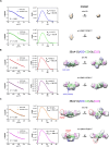
Fig. 3.. Solution SAXS structures of the DNMT3B-3L complexes in the absence or presence of the H3 peptide.
SAXS profiles (black dots) are represented as logarithmic scattering intensities in the left panels. Calculated profiles are displayed as solid-colored lines, with the fitting errors (χ2) shown within the figure. Distance distribution functions [P(r)/I(0)] are shown in the middle panels with radii of gyration (Rg) and maximum diameters (Dmax) shown within the figures. SAXS envelopes of (A) the PWWP domain, (B) 3BΔ412-3L, and (C) 3BΔ214-3L were determined using GASBOR (dashed circles mark the possible location of the PWWP domain). Rigid domains were used to fit into the SAXS envelope: the PWWP domain (this study; PDB: 8ZLK) and the 3BΔ412-3L structural model [built on the basis of the structures of the DNMT3A-3L complex in autoinhibitory and active conformations (20)]. The ADD and PWWP domains are substantially shifted in the 3BΔ412-3L and 3BΔ214-3L complexes in the presence of the H3K4me0K36me0 and H3K4me0K36me3 peptides, respectively.
Fig. 4.
Fig. 4.. The DNMT3B-3L complex is dynamic and adopts multiple conformations.
(A) SAXS profiles of the 3BΔ214-3L complex (black dots) in the absence (left panel) or presence (right panel) of the H3K4me0K36me3 peptide. The SAXS data were analyzed by the EOM and the EOM-simulated curves (solid lines) that were calculated from a group of conformers have been fitted with the SAXS data with low χ2 values. (B) The frequencies of size distribution that resulted from three independent EOM analyses (labeled by Run-1, Run-2, and Run-3) reveal four major conformations with high frequencies. The peaks that correspond to Autoinhibitory-1, Autoinhibitory-2, Activated-1, and Activated-2 conformations are labeled in the figure. (C) Percentage ranges of multiple conformations (Autoinhibitory Form 1, Autoinhibitory Form 2, Activated Form 1, and Activated Form 2) of wild-type 3BΔ214-3L, as well as the two mutant complexes (E515D and R545C), generated from three independent EOM analyses in the absence or presence of the H3K4me0K36me3 peptide are shown.
Fig. 5.
Fig. 5.. The PWWP domain of DNMT3B directly interacts with the ADD domain upon H3 peptide binding.
(A) Structure model of the 3BΔ214-3L complex (SAXS model in Activated Form 2) bound with an H3 peptide (left panel) and structural model of the 3BΔ214-3L-H3 peptide complex bound with DNA (right panel). (B) In the absence of the H3K4me0K36me3 peptide, GST pull-down assays show that the GST-tagged ADD domain failed to pull down the PWWP domain. However, in the presence of the H3K4me0K36me3 peptide, the GST-tagged ADD domain pulled down the PWWP domain. The ADD mutant D470A failed to pull down the PWWP domain in the presence or absence of the H3K4me0K36me3 peptide. The assays were quantified by band densitometry and plotted as the means ± SD for three independent experiments (right panel).
Fig. 6.
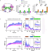
Fig. 6.. Oncogenic R545C mutation enhances DNMT3B’s activity upon interactions with the H3 peptide because of increased flexibility at PWWP and ADD domains.
(A) The locations of the two mutations, E515D and R545C, on the surface of the ADD domain are displayed as ball models in the structure of the 3BΔ214-3L complex in the active conformation. (B) Left panel: relative methyltransferase activities of wild-type 3BΔ412-3L (set to 1) and two corresponding E515D and R545C mutant variants in the absence or presence of the H3K4me0K36me0 peptide. Right panel: relative methyltransferase activities of the wild-type 3BΔ213-3L complex (set to 1) and two E515D and R545C mutant variants in the absence or presence of the H3K4me0K36me3 peptide. Error bars denote SD. Statistical significance (P values) was determined by two-tailed Student’s t test: **P < 0.01, ***P < 0.001, and ****P < 0.0001. (C) Three independent all-atom molecular dynamics stimulations of 3BΔ214-3L-H3 complexes in the active state (Activated Form 2) for wild-type (WT) 3BΔ214-3L and mutated R545C complexes were performed in explicit solvent for 100 ns at 300 and 310 K, respectively. The dynamics of the PWWP and ADD domains were increased in the R545C mutant, which exhibited higher RMSF values in these regions than those of the wild-type 3BΔ214-3L complex. The averaged RMSD (between the initial and simulated structures) profiles and RMSF (for the residues that fluctuate from their original mean positions) profiles from three independent runs are displayed in solid red and blue lines with the error bar representing 1 SD showing in red and blue shades for wild-type and R545C mutant complexes, respectively. The RMSF profile in the loop region between the ADD domain and CD is enlarged at the right upper corner, revealing higher fluctuations in the R545C mutant complex.
Fig. 7.
Fig. 7.. Conformational rearrangement of DNMT3B triggered by H3 peptide binding unleashes its DNA binding and methylation activity.
The structural model of the DNMT3B-3L complex in the absence of the H3K4me0K36me3 peptide shows that it is primarily in the autoinhibitory state. In this autoinhibitory conformation, the ADD domain blocks the DNA binding region in the CD. Binding of the H3K4me0K36me3 peptide to DNMT3B induces a conformational rearrangement of the PWWP and ADD domains to facilitate DNA binding and methylation. In the active state, the PWWP domain concurrently interacts with the H3 peptide and DNA. See also movie S1.

References

    1. Feng Y., Endo M., Sugiyama H., Nucleosomes and epigenetics from a chemical perspective. Chembiochem 22, 595–612 (2021). - PubMed
    1. Zhang W., Xu J., DNA methyltransferases and their roles in tumorigenesis. Biomarker Res. 5, 1 (2017). - PMC - PubMed
    1. Lyko F., The DNA methyltransferase family: A versatile toolkit for epigenetic regulation. Nat. Genet. Rev. 19, 81–92 (2018). - PubMed
    1. Denis H., Ndlovu M. N., Fuks F., Regulation of mammalian DNA methyltransferases: A route to new mechanisms. EMBO Rep. 12, 647–656 (2011). - PMC - PubMed
    1. Laisné M., Gupta N., Kirsh O., Pradhan S., Defossez P.-A., Mechanisms of DNA methyltransferase recruitment in mammals. Genes 9, 617 (2018). - PMC - PubMed

Substances