. 2025 Mar 28;11(13):eadt9354.
doi: 10.1126/sciadv.adt9354. Epub 2025 Mar 26.

Retinal gene therapy for Stargardt disease with dual AAV intein vectors is both safe and effective in large animal models

Affiliations

Retinal gene therapy for Stargardt disease with dual AAV intein vectors is both safe and effective in large animal models

Rita Ferla et al. Sci Adv. .

Abstract

Retinal gene therapy using dual adeno-associated viral (AAV) intein vectors can be applied to genetic forms of blindness caused by mutations in genes with coding sequences that exceed single AAV cargo capacity, such as Stargardt disease (STGD1), the most common inherited macular dystrophy. In view of clinical translation of dual AAV intein vectors, here we set to evaluate both the efficiency and safety of their subretinal administration in relevant large animal models. Accordingly, we have developed the first pig model of STGD1, which we found to accumulate lipofuscin similarly to patients. This accumulation is significantly reduced upon subretinal administration of dual AAV intein vectors whose safety and pharmacodynamics we then tested in nonhuman primates, which showed modest and reversible inflammation as well as high levels of photoreceptor transduction. This bodes well for further clinical translation of dual AAV intein vectors in patients with STGD1 as well as for other blinding diseases that require the delivery of large genes.


Figures

Fig. 1.
Fig. 1.. Characterization of the STGD1 pig model.
(A) Western blot analysis to assess ABCA4 levels in STGD1 (KO) pig retinas derived from different fibroblasts clones. The ID of the clones is indicated above each lane. WT, wild-type. (B) Representative images of retinal cryosections from WT and KO pigs at 10 months of age, showing intense lipofuscin accumulation (red autofluorescence in the RPE layer), in the absence of evident retinal degeneration. RPE, retinal pigment epithelium; ONL, outer nuclear layer; INL, inner nuclear layer. (C) Quantification of lipofuscin accumulation in KO pig retinas via fluorescence microscopy. Lipofuscin autofluorescence was separately evaluated in four regions of the retina, defined according to the known cone density [fig. S3 and (6)]; measurements were done in 8.5-month-old (n = 2; i.e., 63OD and 65OD), 10-month-old (n = 6; i.e., 7OD, 7OS, 14OD, 14OS, 15OD, and 15OS), and 15-month-old (n = 2; i.e., 731OD and 731OS) KO eyes; WT: average lipofuscin levels from n = 6 eyes (i.e., 707OD, 707OS, 850OD, 850OS, 857OD, and 857OS). m.o., month-old. (D) A2E levels in KO pig RPE; measurements were done in 10- to 15-month-old WT (n = 6; i.e., 707OD, 707OS, 850OD, 850OS, 857OD, and 857OS) and KO (n = 8; 7OD, 7OS, 14OD, 14OS, 15OD, 15OS, 731OD, and 731OS) eyes. ***P < 0.001, Welch’s t test. (E) Quantification of ONL thickness in the four retinal regions of KO pig eyes. Measurements were done in 8.5-month-old (n = 2; i.e., 63OD and 65OD), 10–10.5-month-old (n = 8; i.e., 6OD, 7OD, 7OS, 12OD, 14OD, 14OS, 15OD, and 15OS), and 15-month-old (n = 2; i.e., 731OD and 731OS) KO eyes; WT: average levels from n = 6 eyes (i.e., 707OD, 707OS, 850OD, 850OS, 857OD, and 857OS). (F) Measurements of retinal electrical activity via ERG analysis in KO pigs at 2 to 3 years of age. *P < 0.05, Welch’s t test. [(C) to (F)] Data are presented as average ± SEM. Each dot represents a retina.
Fig. 2.
Fig. 2.. AAV8.ABCA4.intein mediated gene therapy in STGD1 pigs.
(A) Western blot (WB) analysis on retinal lysates from both FB-injected WT eyes and from STGD1 pig (KO) eyes either injected with AAV8.ABCA4.intein vectors or with FB. Further details on the areas of the retina used for the WB are provided in fig. S4. The anti-ABCA4 antibody used in the WB reacts with both human (expressed from AAV8.ABCA4.intein vectors; dashed arrow) and swine (i.e., endogenous in WT; straight arrow) ABCA4. (B) Representative images of retinal cryosections from FB-treated WT (top), FB-treated KO (middle), and AAV8.ABCA4.intein injected KO (bottom) eyes. RPE, retinal pigment epithelium; ONL, outer nuclear layer. (C) Quantification of lipofuscin accumulation in the area with highest levels of transduction in AAV8.ABCA4.intein-treated eyes (2 months postinjection), expressed as ratio relative to the same area in contralateral FB-treated eyes. Light gray bars represent average values (± SEM) of measurements taken in the three AAV8.ABCA4.intein-treated KO eyes (pig ID is reported below the graph). The dark gray bar represents average values (± SEM) of measurements taken in the three contralateral FB-treated KO eyes. Dots represent single measurements in each retinal section. Data were analyzed using one-way ANOVA followed by Dunnett’s test, *P < 0.05.
Fig. 3.
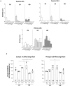
Fig. 3.. Intraocular inflammation and electroretinography (ERG) responses upon AAV8.ABCA4.intein subretinal administration in NHPs.
(A) Aqueous cells, aqueous flare, and vitreous cells were monitored in injected eyes at different time points postinjection. A score (from 1 to 4) was attributed to define the severity of the ocular findings in each eye. White bars: formulation buffer–treated eyes (FB); light gray bars: eyes treated with a low dose of AAV8.ABCA4.intein vectors (LD); dark gray bars: eyes treated with a high dose of AAV8.ABCA4.intein vectors (HD). Bars represent average values ± SEM for each group. Detailed number of eyes analyzed and values measured in each eye at each time point are presented in fig. S6. (B) ERG values in right eyes treated with either an LD or an HD of AAV8.ABCA4.intein vectors are expressed as percentage relative to left FB-treated eyes. Data are presented as means ± SEM. Each dot represents one eye. Pre, analysis performed before injection; w, weeks postinjection. Statistical comparisons were performed using the two-way ANOVA followed by Tukey’s post hoc, *P < 0.05. Further details on statistical analysis, including exact P values, can be found in table S7.
Fig. 4.
Fig. 4.. High levels of retinal transduction following subretinal delivery of AAV8.ABCA4.intein vectors in NHPs.
(A) Representative pictures of retinal sections analyzed by BaseScope from NHP eyes at 13 weeks postinjection of either an LD (n = 3) or HD (n = 3) of AAV8.ABCA4.intein vectors or the FB (n = 6). Probes annealing to either the Npu N-intein (dark green dots) or C-intein (red dots) of human.ABCA4.intein mRNAs were used to assess transduction. ONL, outer nuclear layer. (B) ABCA4 protein levels were measured by Simple Western analysis in two retina punches [one superior temporal (black dot) and one superior nasal (gray dot) punch] collected from the injected bleb area of eyes receiving either a subretinal injection of AAV8.ABCA4.intein at the LD (n = 2) or HD (n = 2) or FB (n = 1) as control. ABCA4 protein levels were normalized to the endogenous control (PDE6B) and then expressed as percentage (%) of ABCA4 protein levels in AAV8.ABCA4.intein-treated versus FB-treated areas. Columns represent mean values for group of treatment, whereas dots represent value of each retina punch. Data are reported as means ± SEM.

References

    1. Molday R. S., Garces F. A., Scortecci J. F., Molday L. L., Structure and function of ABCA4 and its role in the visual cycle and Stargardt macular degeneration. Prog. Retin. Eye Res. 89, 101036 (2022). - PubMed
    1. Cowan C. S., Renner M., De Gennaro M., Gross-Scherf B., Goldblum D., Hou Y., Munz M., Rodrigues T. M., Krol J., Szikra T., Cuttat R., Waldt A., Papasaikas P., Diggelmann R., Patino-Alvarez C. P., Galliker P., Spirig S. E., Pavlinic D., Gerber-Hollbach N., Schuierer S., Srdanovic A., Balogh M., Panero R., Kusnyerik A., Szabo A., Stadler M. B., Orgül S., Picelli S., Hasler P. W., Hierlemann A., Scholl H. P. N., Roma G., Nigsch F., Roska B., Cell types of the human retina and its organoids at single-cell resolution. Cell 182, 1623–1640.e34 (2020). - PMC - PubMed
    1. Lenis T. L., Hu J., Ng S. Y., Jiang Z., Sarfare S., Lloyd M. B., Esposito N. J., Samuel W., Jaworski C., Bok D., Finnemann S. C., Radeke M. J., Redmond T. M., Travis G. H., Radu R. A., Expression of ABCA4 in the retinal pigment epithelium and its implications for Stargardt macular degeneration. Proc. Natl. Acad. Sci. U.S.A. 115, E11120–E11127 (2018). - PMC - PubMed
    1. Simons E. J., Trapani I., The opportunities and challenges of gene therapy for treatment of inherited forms of vision and hearing loss. Hum. Gene Ther. 34, 808–820 (2023). - PubMed
    1. Tornabene P., Trapani I., Minopoli R., Centrulo M., Lupo M., de Simone S., Tiberi P., Dell’Aquila F., Marrocco E., Iodice C., Iuliano A., Gesualdo C., Rossi S., Giaquinto L., Albert S., Hoyng C. B., Polishchuk E., Cremers F. P. M., Surace E. M., Simonelli F., De Matteis M. A., Polishchuk R., Auricchio A., Intein-mediated protein trans-splicing expands adeno-associated virus transfer capacity in the retina. Sci. Transl. Med. 11, eaav4523 (2019). - PMC - PubMed