. 2025 Mar 26;13(3):e010791.
doi: 10.1136/jitc-2024-010791.

Oxaliplatin, ATR inhibitor and anti-PD-1 antibody combination therapy controls colon carcinoma growth, induces local and systemic changes in the immune compartment, and protects against tumor rechallenge in mice

Affiliations

Oxaliplatin, ATR inhibitor and anti-PD-1 antibody combination therapy controls colon carcinoma growth, induces local and systemic changes in the immune compartment, and protects against tumor rechallenge in mice

Alexandra Fauvre et al. J Immunother Cancer. .

Abstract

Background: Colorectal cancer (CRC) is the third most common cancer type and one of the leading causes of cancer-related death worldwide. The treatment of advanced metastatic CRC relies on classical chemotherapy combinations (5-fluorouracil, oxaliplatin or irinotecan). However, their use is limited by the emergence of resistance mechanisms, including to oxaliplatin. In this context, we recently showed that the combination of oxaliplatin and ataxia telangiectasia and Rad3-related protein inhibition (VE-822) is synergistic and may have a potential therapeutic effect in metastatic CRC management.

Methods: In this study, we investigated the role of the VE-822+oxaliplatin (Vox) combination on the immune response and its potential synergy with an anti-programmed-cell Death receptor-1 (PD-1) antibody. We used cell lines and organoids from metastatic CRC to investigate in vitro Vox efficacy and orthotopic syngeneic mouse models of metastatic CRC to assess the efficacy of Vox+anti-PD-1 antibody and identify the involved immune cells.

Results: The Vox+anti-PD-1 antibody combination completely cured tumor-bearing mice and protected them from a rechallenge. Vox was associated with a reduction of tumor-infiltrated neutrophils, CD206+ macrophages and regulatory T cells. Vox also induced a deep depletion of blood neutrophils. The increased bone marrow granulopoiesis failed to compensate for the Vox-mediated mature neutrophil depletion. Neutrophil depletion using a mouse recombinant anti-Ly6G antibody partially mimicked the Vox effect on the tumor microenvironment, but to a lower extent compared with the Vox+anti-PD-1 antibody combination. Vox, but not neutrophil depletion, led to the emergence of an Ly6C+ PD-1+ CD8+ T-cell population in the blood and spleen of tumor-harboring mice. These cells were proliferating, and expressed IFN-γ, CD62L, CXCR3 and Eomes. Moreover, the proportion of tumor antigen-specific T cells and of CD122+ BCL6+ T cells, which shared phenotypic characteristics with stem-like CD8+ T cells, was increased in treated mice.

Conclusions: Our work strongly suggests that the Vox+anti-PD-1 antibody combination might significantly improve survival in patients with metastatic and treatment-refractory CRC by acting both on cancer cells and CD8+ T cells.

Keywords: Colorectal Cancer; Immune Checkpoint Inhibitor; Neutropenia; T cell.


Conflict of interest statement

Competing interests: None declared.

Figures

Figure 1
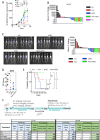
Figure 1. Vox, but not oxaliplatin and VE-822 alone, reduces tumoroid growth. (A) Representative images (left) of patient-derived tumoroids showing viable cells (green) after 14 days of culture in the presence of oxaliplatin (Ox; 0.3 µM), VE-822 (Ve; 1 µM) or Vox (Ox: 0.3 µM+Ve: 1 µM) for 7 days (n=8 patients). Histogram (right) showing the percentage of live cells (green) compared with control (not treated, NT). *p<0.05 (Mann-Whitney test). (B and C): (B) Western blots showing PD-L1 expression in HCT116 and HCT116-R1 cells NT or incubated with increasing concentrations of oxaliplatin (from 12.5 µM to 100 µM) or (C) with oxaliplatin (25 µM), VE-822 (1 µM) or Vox (Ox: 25 µM+Ve: 1 µM) for 24 hours. PD-1, programmed-cell death ligand-1; Vox, VE-822+oxaliplatin.
Figure 2
Figure 2. The Vox and anti-PD-1 immunotherapy combination cures mice harboring colorectal cancer-PM. (A) Curves show tumor growth in mice bearing subcutaneous MC38 tumors treated with PBS (NT, n=7), oxaliplatin (Ox; 5 mg/kg once every 2 weeks; n=8), VE-822 (Ve; 40 mg/kg two times a week; n=8) or Vox (same modality as for each drug alone; n=8) for 4 weeks. Mixed model statistic test. (B) Histogram showing the percentage of luciferase signal intensity change in GFP-Luc-MC38 PM-harboring mice treated with PBS (Ctr, n=7), Vox (Ve+Ox, n=6), anti-PD-1 antibody (100 mg two times a week, n=6), VE-822 (Ve; 40 mg/kg, n=2), or Vox+anti-PD-1 antibody (n=6). (C) Representative images (left) of luciferase signal intensity in mice harboring GFP-Luc-CT26 PM (control NT n=9 and Vox+anti-PD-1 antibody n=9) at day 22 after treatment initiation and histogram (right) showing the percentage of luciferase signal intensity change (Ctr n=9, Vox n=6, anti-PD-1 antibody n=6, and Vox+anti-PD-1 antibody n=6). (D) Plot showing the PCI scores of mice harboring GFP-Luc-CT26 PM treated as in C and sacrificed at week 3–8 after treatment initiation. (E) Kaplan-Meier curves showing survival of mice harboring GFP-Luc-CT26 PM treated as in C. (F) Tables showing the number and percentage of tumor-free mice after the first challenge and the number and proportion of cured mice that did not develop tumor lesions following the rechallenge in the indicated conditions. *p<0.05; **p<0.01; ***p<0.001; ****p<0.0001; Ctr, control; ns, not significant; NT, not treated; PBS, phosphate-buffered saline; PCI, peritoneal cancer index; PD-1, programmed-cell death receptor-1; PM, peritoneal metastases; Vox, VE-822+oxaliplatin.
Figure 3
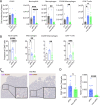
Figure 3. The Vox+anti-PD-1 antibody combination reduces neutrophil, CD206+ macrophages and regulatory T cell infiltration in PM nodules. (A) Number per mg of tumor of the indicated immune cell populations measured by quantitative flow cytometry in GFP-Luc-CT26 PM samples harvested from untreated controls (Ctr n=7) and mice treated with the anti-PD-1 antibody (n=8) alone and with Vox (n=9) for 1 week. (B) Histograms showing the percentage among all tumor-infiltrated CD45+ immune cells of the indicated immune cell populations from the same experiment described in A. (C) Representative images of CD8 (purple) and FOXP3 (brown) double staining in GFP-Luc-CT26 PM samples from mice treated with the anti-PD-1 alone (n=15) and with Vox (n=13). (D) Histograms showing the ratio of CD8+ T cells and FOXP3+ cells over the total cell number per tumor from the IHC staining images. *p=0.05; **p=0.01; ***p=0.001; ****p=0.0001; ns, not significant (one-way analysis of variance for A and B and Student’s t-test for D). aPD-1, anti-programmed-cell death receptor-1; Ctr, control; IHC, immunohistochemistry; PM, peritoneal metastases; Vox, VE-822+oxaliplatin.
Figure 4
Figure 4. Vox reduces neutrophils and CD206+ macrophages and drives the expression of antitumor immune response genes in GFP-Luc-CT26 PM lesions. (A) Number per mg of tumor of CD45+ immune cells measured by quantitative flow cytometry in GFP-Luc-CT26 PM samples from untreated control (Ctr n=11) or mice treated with oxaliplatin (Ox n=12), VE-822 (Ve n=12), or Vox (n=12) for 1 week. (B, C and D) Heatmap (B) and histograms (C–D) showing the number per mg of tumor of the indicated immune cell populations measured in the same tumor samples described in (A). (E) Percentages of the indicated immune cell populations relative to all tumor-infiltrated immune cells (CD45+) obtained using data from two independent experiments (filled circles: same experiment as in A; empty circles: another experiment where Ctr and Vox-treated PM samples were compared) (Ctr n=25, Ox n=12, Ve n=12, and Vox n=19). (F) Histograms showing the relative mRNA expression in sorted cancer cells (GFP+CD45DAPI), total immune cells (CD45+DAPI), macrophages (CD45+CD11b+Ly-6GF4/80+DAPI) and neutrophils (CD45+CD11b+Ly-6G+F4/80DAPI) from GFP-Luc-CT26 PM lesions harvested from the control group and from mice treated with Vox for 1 week. Each measurement was obtained from a pool of three mice, n=5 pools per condition. *p=0.05; **p=0.01; ***p=0001; ****p=0.0001; ns, not significant (one-way analysis of variance). Ctr, control; GFP, green fluorescent protein; mRNA, messenger RNA; PM, peritoneal metastases; Vox, VE-822+oxaliplatin.
Figure 5
Figure 5. Vox alters neutrophil biogenesis, but neutrophil depletion does not recapitulate Vox antitumor effect. (A) Number per mL of blood of the indicated immune cell populations measured by quantitative flow cytometry in GFP-Luc-CT26 PM mice that were not treated (Ctr n=5) or treated with Vox (n=6), anti-PD-1 antibody (n=6), or Vox+anti-PD-1 antibody (n=6) for 1 week. (B) Number per femur of hematopoietic stem cells (LSK), immature lineage-negative c-Kit+ (LK) cells, granulocyte/monocyte progenitors (GMP), CD11b+Gr-1low and differentiated CD11b+Gr-1high neutrophils from the same mice as in A. (C) Ki67 expression in GMPs and differentiated neutrophils (CD11b+Gr-1+) in the control condition (Ctr n=10) or after Vox treatment (n=10) in mice bearing GFP-Luc-CT26 PM. (D) Total CD45+ cells and Ly-6C-CD11b+ neutrophils by quantitative flow cytometry in GFP-Luc-CT26 PM lesions from mice treated with a control antibody (Ctr) or 150 mg of mouse recombinant anti-Ly-6G antibody (n=6 per condition). (E and F) Cell number per mg of tumor tissue (E) and percentage relative to all tumor-infiltrated immune cells (F) of the indicated immune cell populations from the same samples as in D. *p=0.05; **p=0.01; ***p=0.001; ****p=0.0001 (one-way analysis of variance). (G) Kaplan-Meier survival curves of mice treated with isotype IgG control (not treated, n=8), anti-PD-1 antibody plus isotype IgG control (PD-1+IsoT, n=8), anti-PD-1 antibody+anti-Ly-6G antibody (n=8) and anti-PD-1 antibody+Vox (n=8); ***p=0.001. aPD-1, anti-programmed-cell death receptor-1; Ctr, control; GFP, green fluorescent protein; PM, peritoneal metastases; Vox, VE-822+oxaliplatin.
Figure 6
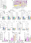
Figure 6. Vox treatment leads to the emergence of Ly-6C+PD-1+ CD8+ T cells in blood and spleen. (A and B) Number per mL of blood (A) and percentage relative to immune cells (B) of the indicated CD8+ T-cell populations from GFP-Luc-CT26 PM mice untreated (Ctr n=5), or treated with Vox (n=6), anti-PD-1 antibody (n=6) and Vox+anti-PD-1 antibody (n=5). (C) Number per spleen of CD8+ T cells with the indicated phenotypes from GFP-Luc-CT26 PM mice untreated (Ctr n=7), or treated with the anti-PD-1 antibody (n=16) or with Vox+anti-PD-1 antibody (n=10). (D) Histogram showing the percentage of Ki67+ cells among the indicated CD8+ T-cell subpopulations from GFP-Luc-CT26 PM mice untreated (Ctr n=6) or treated with the anti-PD-1 antibody (n=6) or Vox+anti-PD-1 antibody (n=5). (E) Relative messenger RNA expression of the indicated genes in the indicated CD8+ T-cell subpopulations purified from the spleen of Vox-treated GFP-Luc-CT26 PM mice (n=5). (F) CXCR3, Eomes and CD62L expression in CD8+ T cells (flow cytometry analysis) from the spleen of Vox-treated GFP-Luc-CT26 PM mice. *p=0.05; **p=0.01; ***p=0.001; ****p=0.0001; ns, not significant (one-way analysis of variance). aPD-1, anti-programmed-cell death receptor-1; Ctr, control; GFP, green fluorescent protein; PM, peritoneal metastases; Vox, VE-822+oxaliplatin.
Figure 7
Figure 7. The Ly-6C+PD-1+ CD8+ T-cell population contains a strong proportion of tumor antigen-specific T cells. (A) Histogram showing the percentage of BCL6+CD122+ cells among PD-1+Ly-6C+ T cells (black) and PD-1+Ly-6C CD8+ T cells (white) from the spleen of GFP-Luc-CT26 PM mice treated with the anti-PD-1 antibody (n=8) or Vox+anti-PD-1 antibody (n=11). (B) Plots showing the gating strategy to identify GFP-specific T-cell receptor-positive cells after Dextramer staining and Ly-6C+ and PD-1+ cells among them in the spleen of Vox+anti-PD-1 antibody-treated GFP-Luc-CT26 PM mice. (C) Pie chart (top) and histogram (bottom) showing the distribution of dextran-positive (right) and dextran-negative (left) cells in the different CD8+ T-cell subpopulations from the spleen of Vox+anti-PD-1 antibody-treated GFP-Luc-CT26 PM mice (n=5). *p=0.05; **p=0.01 (one-way analysis of variance). PD-1, anti-programmed-cell death receptor-1; GFP, green fluorescent protein; PM, peritoneal metastases; Vox, VE-822+oxaliplatin.

References

    1. Sung H, Ferlay J, Siegel RL, et al. Global Cancer Statistics 2020: GLOBOCAN Estimates of Incidence and Mortality Worldwide for 36 Cancers in 185 Countries. CA Cancer J Clin. 2021;71:209–49. doi: 10.3322/caac.21660. - DOI - PubMed
    1. Jooste V, Lepage C, Manfredi S, et al. Trends in incidence of infrequent and frequent synchronous metastases from colorectal cancer. Dig Liver Dis. 2025;57:83–8. doi: 10.1016/j.dld.2024.06.018. - DOI - PubMed
    1. Franko J, Shi Q, Meyers JP, et al. Prognosis of patients with peritoneal metastatic colorectal cancer given systemic therapy: an analysis of individual patient data from prospective randomised trials from the Analysis and Research in Cancers of the Digestive System (ARCAD) database. Lancet Oncol. 2016;17:1709–19. doi: 10.1016/S1470-2045(16)30500-9. - DOI - PubMed
    1. Van Cutsem E, Nordlinger B, Cervantes A. Advanced colorectal cancer: ESMO Clinical Practice Guidelines for treatment. Ann Oncol. 2010;21:v93–7. doi: 10.1093/annonc/mdq222. - DOI - PubMed
    1. Golshani G, Zhang Y. Advances in immunotherapy for colorectal cancer: a review. Therap Adv Gastroenterol . 2020;13:1756284820917527. doi: 10.1177/1756284820917527. - DOI - PMC - PubMed

MeSH terms

Substances

LinkOut - more resources