. 2025 Mar;10(3):323-341.
doi: 10.1016/j.jacbts.2024.10.007. Epub 2024 Dec 18.

Homeostatic Role of Decorin in Right Ventricular Pressure Overload and Pulmonary Hypertension Induced Remodeling

Affiliations

Homeostatic Role of Decorin in Right Ventricular Pressure Overload and Pulmonary Hypertension Induced Remodeling

Yohei Akazawa et al. JACC Basic Transl Sci. 2025 Mar.

Abstract

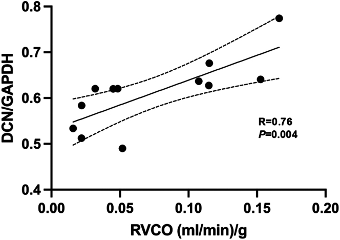
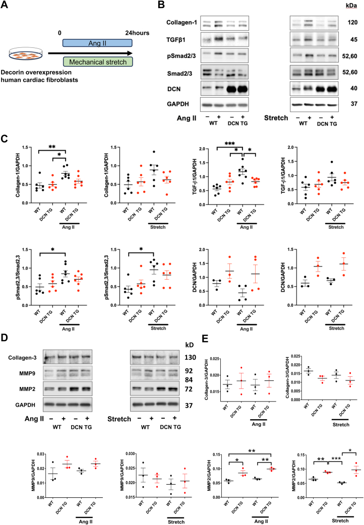
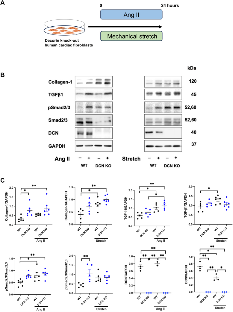
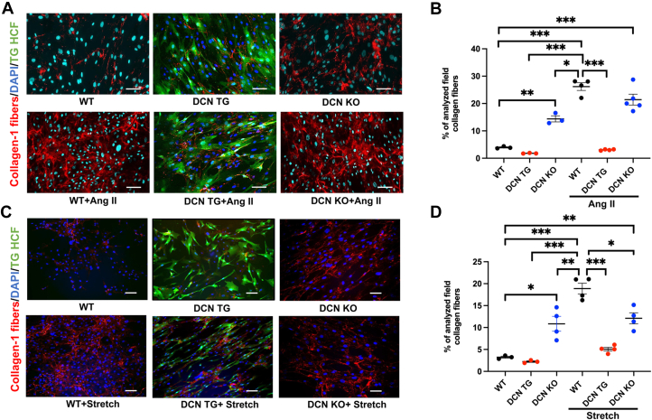
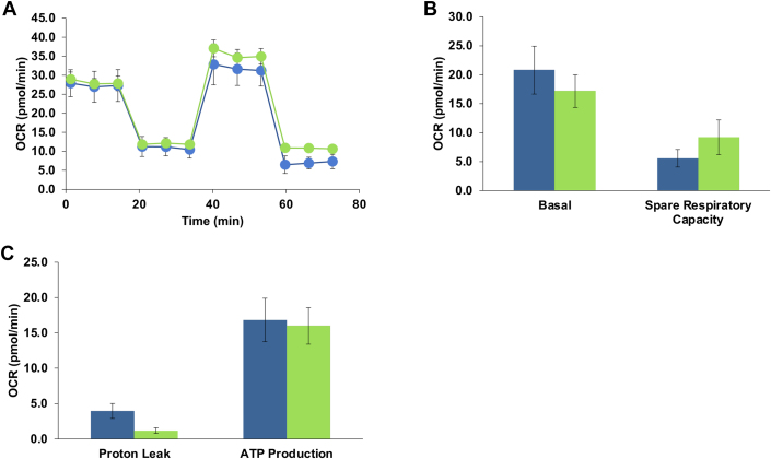
Right ventricular (RV) pressure loading induces RV profibrotic signaling and fibrosis associated with RV dysfunction. RV decorin protein levels are decreased in patients with chronic RV pressure loading. RV decorin protein levels are also decreased in 4 animal models of mechanical RV pressure loading and pulmonary arterial hypertension. Human cardiac fibroblasts overexpressing decorin show diminished collagen-1 secretion in response to mechanical or chemical profibrotic stress while decorin knockout human cardiac fibroblasts show increased collagen-1 secretion in response to stress. Downregulation of decorin may play a key role in upregulating transforming growth factor-β1 profibrotic signaling and fibrosis that contribute to RV dysfunction in RV pressure loading.

Keywords: cardiac fibrosis; decorin; pulmonary hypertension; right ventricular dysfunction; right ventricular pressure loading; transforming growth factor-β1.


Conflict of interest statement

Funding Support and Author Disclosures This work was supported in part by the Heart and Stroke Foundation of Canada and by the Canadian Institutes of Health Research (FRN 16226). Dr Connelly holds the Keenan Chair for Research Leadership, Keenan Research Centre for Biomedical Science, Toronto. All other authors have reported they have no relationships relevant to the contents of this paper to disclose.

Figures

None
Graphical abstract
Figure 1
Figure 1
Decorin Overexpression in CRISPR/Cas Genetically Edited Human Cardiac Fibroblasts (A) Construct expressing decorin and green fluorescent protein (GFP) concurrently. LHA and RHA refer to the left and right homology arms that target the insertion of the construct into DSB created by Cas9/single guide RNA complex in the C-C chemokine receptor type 5 (CCR5) locus. (B) Approximately 30% of the edited cardiac fibroblasts were GFP+ after 10 days. These cells were enriched using fluorescence-activated cell sorting (FACS). On day 14, >85% of sorted cells were GFP+. FSC-A = forward scatter area.
Figure 2
Figure 2
Downregulation of DCN Levels in Animal Models of RV Pressure Loading Decorin (DCN) was downregulated in the right ventricular (RV) free-wall tissue in 4 animal models of chronic RV pressure loading. (A) Representative Western blots detecting indicated DCN and their quantitative assessments in the RV of sham and (a) RV pressure-loading models of rabbit pulmonary artery banding (PAB), (b) rat monocrotaline (MCT), (c) rat sugen plus hypoxia (SuHx), and (d) rat PAB compared with sham controls. Data from n = 3-7 animals per group are presented as mean ± SEM. Unpaired Student’s t-test was performed for statistical analysis. (B) Representative Picrosirius red (PSR) staining of rat hearts (a) rat sham, (b) rat MCT, (c) rat SuHx, and (d) rat PAB. PSR staining demonstrates a substantial increase in cardiac fibrosis in all pulmonary arterial hypertension models (B). (C) Immunostaining detection of DCN-positive labeling (brown) in (a) rat sham, (b) rat MCT, (c) rat SuHx, and (d) rat PAB. Scale Bar = 40 μm. (D) Quantification of immunodetection of RV free-wall DCN levels by stereology in rat SuHx pulmonary hypertension (PH) and rat PAB models. Data from n = 7-8 animals per group are presented as mean ± SEM. Unpaired Student’s t-test was performed for statistical analysis. (E) Representative immunofluorescence staining for DCN expression in various cardiac cell types of sham rat RV tissue. (a) Positive labeling of DCN (red) and vimentin (green), merged show all fibroblasts colocalized with DCN. (b) Positive labeling of DCN (green) and CD31 (red), arrow shows CD31 colocalizing with DCN. (c) Positive labeling of DCN (green) and α-smooth muscle actin (SMA) (red), arrow shows α-SMA colocalizing with DCN. Scale bar = 40 μm. (F) Published RNA sequencing data for DCN expression in various cardiac cell types. (A.) DC expression in cells from the "mouse integrated" dataset in CLARA. (B) DCN expression in cells from the "human heart" dataset in CLARA. (C.) DCN expression in cardiac fibroblast data published by Dewar et al. ∗P < 0.05, ∗∗P < 0.01, and ∗∗∗P < 0.001. GAPDH = glyceraldehyde 3-phosphate dehydrogenase.
Figure 3
Figure 3
Reduced DCN Levels in RV of Patients With Severe RV Pressure Loading DCN expression levels in right ventricular (RV) myocardium are lower in tetralogy of Fallot patients with severe RV pressure overloading (right ventricular outflow tract pressure gradient >70 mm Hg) compared with those of patients with moderate (30-70 mm Hg) and mild (<30 mm Hg) RV pressure loading. Abbreviations as in Figure 2.
Figure 4
Figure 4
Correlation Between RV DCN Expression Levels and Cardiac Output in Rats With RV Pressure Loading Correlation between DCN expression levels and cardiac output indexed for body weight derived from catheter measurements in rats with monocrotaline (n = 5), sugen plus hypoxia (n = 2), and pulmonary artery banding (n = 5). Linear regression analysis was performed for statistical analysis (Pearson’s correlation coefficient). RVCO = right ventricular cardiac output; other abbreviations as in Figure 2.
Figure 5
Figure 5
Amelioration of Fibrosis Signaling and Collagen-1, MMP Secretion in Human Cardiac Fibroblasts Overexpressing DCN CRISPR/Cas genetically manipulated human cardiac fibroblasts overexpressing DCN show ameliorated fibrosis signaling and collagen secretion in response to stress. Angiotensin II (Ang II) stimulation of human cardiac fibroblasts upregulates collagen-1 and transforming growth factor (TGF)-β1 expression levels in wild-type (WT) but not in cardiac fibroblasts overexpressing DCN. Ang II and mechanical stretch each increase pSmad2/3/Smad2/3 significantly in WT but not in cardiac fibroblasts overexpressing DCN. Matrix metalloproteinase (MMP)-2 expression level was significantly increased in DCN overexpressing fibroblasts in both untreated and treated cells. There was no significant change in collagen-3 and MMP-9 expression. (A) Schematic of the experimental design. (B) Representative Western blots detecting collagen-1, TGF-β1, pSmad2/3/Smad2/3, DCN, and GAPDH in DCN overexpressing transgenic human cardiac fibroblasts (DCN TG) and nonmodified control human cardiac fibroblasts (WT). (C) Quantification of collagen-1, TGF-β1, pSmad2/3, Smad2/3, DCN in WT, and DCN TG. ∗P < 0.05, ∗∗P < 0.01, and ∗∗∗P < 0.001. (D) Representative Western blots detecting collagen-3, MMP-2, and MMP-9 in DCN TG human cardiac fibroblast and nonmodified control human cardiac fibroblasts (WT). (E) Quantification of collagen-III, MMP-2, and MMP-9 in WT and DCN TG. ∗P < 0.05, ∗∗P < 0.01, ∗∗∗P < 0.001. Data from n = 3-7 samples per group are presented as mean ± SEM. One-way analysis of variance with Tukey’s post hoc test was performed for statistical analysis. Abbreviations as in Figure 2.
Figure 6
Figure 6
Upregulation of Profibrotic Signaling and Collagen-1 Secretion in Human Cardiac Fibroblasts and in Genetically Edited DCN KO Human Cardiac Fibroblasts Ang II stimulation upregulates collagen-1, TGF-β1, pSmad2/3/Smad2/3, and DCN expression levels in human cardiac fibroblasts and in genetically edited DCN knockout (DCN KO) human cardiac fibroblasts. (A) Schematic of the experimental design. (B) Representative Western blots detecting collagen-1, TGF-β1, pSmad2/3, Smad2/3, and GAPDH in DCN KO human cardiac fibroblasts and control human cardiac fibroblasts (WT). (C) Quantitative assessment of collagen-1, TGF-β1, pSmad2/3, Smad2/ in control and DCN KO human cardiac fibroblasts. ∗P < 0.05, ∗∗P < 0.01. Data from n = 5-6 samples per group are presented as mean ± SEM. One-way analysis of variance with Tukey’s post hoc test was performed for statistical analysis. Abbreviations as in Figures 2 and 5.
Figure 7
Figure 7
Immunodetection of Collagen-1 Levels in Genetically Modified Human Cardiac Fibroblasts Where DCN Is Overexpressed or Knocked Out Representative micrographs of cultured human cardiac fibroblasts immune-stained with anti-collagen-1 antibody further confirms effects of DCN overexpression (DCN TG) or knockout (DCN KO) on collagen deposition. Immunodetection of collagen-1 deposition (red) with (A) Ang II treatment and (C) mechanical stretch. Bars = 40x. Blue depicts nuclear staining by 4ʹ,6-diamidino-2-phenylindole (DAPI). Green depicts DCN expression. (B, D) Following Ang II stimulation and mechanical stretch, immune-detected collagen-1 fiber production increased in control and DCN KO human cardiac fibroblasts vs a smaller increase in DCN overexpressing cells (DCN TG). ∗P < 0.05, ∗∗P < 0.01, and ∗∗∗P < 0.001. Data from n = 3-5 samples per group are presented as mean ± SEM and analyzed using one-way analysis of variance with Tukey’s post hoc test performed for statistical analysis. Abbreviations as in Figures 2, 5, and 6.
Figure 8
Figure 8
OCR in Cultured Right Ventricular Cardiac Fibroblasts Demonstrate differences in mitochondrial function as compared with control cardiac fibroblast at baseline (nontreated control) and after exposure to decorin (5 nM) for 24 hours. Oxygen consumption rates (OCR, in pmole/min) were measured after addition of oligomycin (Oligo, 2 μM), FCCP (2 nM), and rotenone and antimycin A (1 μM each) and basal respiration, Oligo (proton leak), FCCP (maximal respiration), spare respiratory capacity, non-mitochondrial oxygen, ATP production, coupling efficiency, and spare respiratory capacity were calculated and expressed per 3 × 104 cells as shown in (A) mitochondrial respiration, (B) mitochondrial respiration profile, and (C) basal respiration, spare respiratory capacity, proton leak respiration, and ATP-linked respiration.

References

    1. van der Bruggen C.E.E., Tedford R.J., Handoko M.L., van der Velden J., de Man F.S. RV pressure overload: from hypertrophy to failure. Cardiovasc Res. 2017;113:1423–1432. - PubMed
    1. Andersen S., Nielsen-Kudsk J.E., Vonk Noordegraaf A., de Man F.S. Right ventricular fibrosis. Circulation. 2019;139:269–285. - PubMed
    1. Freed B.H., Gomberg-Maitland M., Chandra S., et al. Late gadolinium enhancement cardiovascular magnetic resonance predicts clinical worsening in patients with pulmonary hypertension. J Cardiovasc Magn Reson. 2012;14:11. - PMC - PubMed
    1. Babu-Narayan S.V., Kilner P.J., Li W., et al. Ventricular fibrosis suggested by cardiovascular magnetic resonance in adults with repaired tetralogy of fallot and its relationship to adverse markers of clinical outcome. Circulation. 2006;113:405–413. - PubMed
    1. Yamamura K., Yuen D., Hickey E.J., et al. Right ventricular fibrosis is associated with cardiac remodeling after pulmonary valve replacement. Heart. 2019;105:855–863. - PubMed

LinkOut - more resources