

Nasal cytological evidence of chronic inflammation in the olfactory cleft in post-viral olfactory dysfunction
- PMID: 40140006
- PMCID: PMC12055885
- DOI: 10.1007/s00405-025-09302-2
Nasal cytological evidence of chronic inflammation in the olfactory cleft in post-viral olfactory dysfunction
Abstract
Purpose: This study investigated nasal cytological alterations in patients with persistent post-viral olfactory dysfunction. The primary objective was to evaluate the role of immune dysregulation and chronic local inflammation within the nasal mucosa in sustaining long-term olfactory impairment.
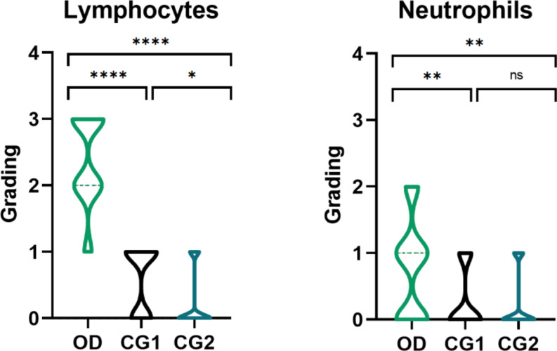
Methods: An observational case-control study was conducted at the Otorhinolaryngology Department of the University of Rome Tor Vergata. Thirty-six patients with persistent olfactory dysfunction were compared to two control groups: one comprised subjects recovered from SARS-CoV-2 infection without olfactory impairment, and the other included individuals without a history of COVID-19 or olfactory dysfunction. Psychophysical olfactory function was assessed using the TDI (Threshold, Discrimination, and Identification) test. Nasal cytology samples were obtained via nasal brushing at the level of the olfactory cleft and stained using the May-Grunwald-Giemsa technique. Cellular alterations were evaluated using a semiquantitative grading system.
Results: Patients with persistent olfactory dysfunction exhibited increased lymphocytes and neutrophils compared to both control groups, indicating ongoing local inflammation. Ciliocytophthoria was notably present in a significant portion of the olfactory dysfunction group, while absent or minimally present in controls. Eosinophils and mast cells were rare across all groups.
Conclusion: Persistent post-viral olfactory dysfunction is associated with sustained immune activation and epithelial damage localized to the olfactory cleft. Elevated lymphocytes, neutrophils, and ciliocytophthoria emphasize the role of chronic inflammation in the pathogenesis of prolonged olfactory deficits. These findings highlight the potential utility of targeted therapies to modulate immune responses and promote olfactory recovery in affected patients.
Keywords: Ciliocytophthoria; Long COVID; Lymphocytes; Nasal cytology; Olfactory dysfunction; Post-Viral.
© 2025. The Author(s).
Conflict of interest statement
Declarations. Ethical approval: The research was conducted in accordance with the principles of the Helsinki Declaration. The study protocol was approved by the Ethical Committee of Policlinico Tor Vergata. Informed consent: Informed consent was obtained from all individual participants included in the study. Conflict of interest: The authors have no relevant financial or non-financial interests to disclose.
Figures

Similar articles
-
Olfactory cleft brushing: A minimally invasive tool for biomarker analysis in rhinology.Am J Otolaryngol. 2025 Jan-Feb;46(1):104589. doi: 10.1016/j.amjoto.2024.104589. Epub 2024 Dec 21. Am J Otolaryngol. 2025. PMID: 39721256
-
Post-viral effects of COVID-19 in the olfactory system and their implications.Lancet Neurol. 2021 Sep;20(9):753-761. doi: 10.1016/S1474-4422(21)00182-4. Epub 2021 Jul 30. Lancet Neurol. 2021. PMID: 34339626 Free PMC article. Review.
-
Autoantibody profiles assessment in individuals with persistent olfactory impairment following SARS-CoV-2 infection.Int Immunopharmacol. 2024 Mar 10;129:111599. doi: 10.1016/j.intimp.2024.111599. Epub 2024 Feb 7. Int Immunopharmacol. 2024. PMID: 38330796
-
Injection of Platelet Rich Plasma in the Olfactory Cleft for COVID-19 Patients With Persistent Olfactory Dysfunction: Description of the Technique.Ear Nose Throat J. 2024 Jun;103(1_suppl):115S-119S. doi: 10.1177/01455613221124773. Epub 2022 Oct 11. Ear Nose Throat J. 2024. PMID: 36219733 Free PMC article.
-
COVID-19 and olfactory dysfunction: a looming wave of dementia?J Neurophysiol. 2022 Aug 1;128(2):436-444. doi: 10.1152/jn.00255.2022. Epub 2022 Jul 27. J Neurophysiol. 2022. PMID: 35894511 Free PMC article. Review.
References
Publication types
MeSH terms
LinkOut - more resources
Full Text Sources
Medical
Miscellaneous
