

Structure of the lens MP20 mediated adhesive junction
- PMID: 40140346
- PMCID: PMC11947226
- DOI: 10.1038/s41467-025-57903-6
Structure of the lens MP20 mediated adhesive junction
Abstract
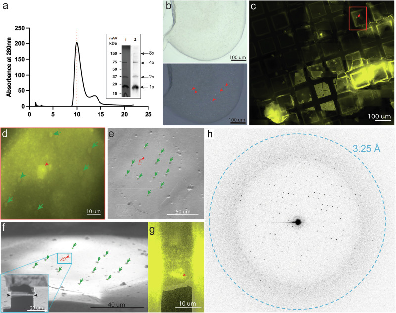
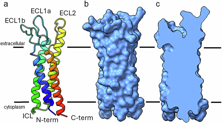
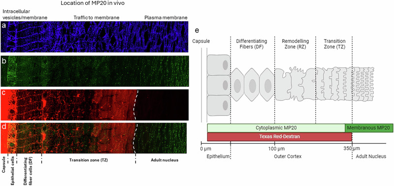
Human lens fiber membrane intrinsic protein MP20 is the second most abundant membrane protein of the human eye lens. Despite decades of effort its structure and function remained elusive. Here, we determined the MicroED structure of full-length human MP20 in lipidic-cubic phase to a resolution of 3.5 Å. MP20 forms tetramers each of which contain 4 transmembrane α-helices that are packed against one another forming a helical bundle. We find that each MP20 tetramer formed adhesive interactions with an opposing tetramer in a head-to-head fashion. Investigation of MP20 localization in human lenses indicate that in young fiber cells MP20 is initially localized to the cytoplasm in differentiating fiber cells but upon fiber cell maturation is inserted into the plasma membrane, correlating with the restriction of the diffusion of extracellular tracers into the lens. Together these results suggest that MP20 forms lens thin junctions in vivo, confirming its role as a structural protein in the human eye lens essential for its optical transparency.
© 2025. The Author(s).
Conflict of interest statement
Competing interests: The authors declare no competing interests.
Figures

Update of
-
Structure of the lens MP20 mediated adhesive junction.bioRxiv [Preprint]. 2024 May 13:2024.05.13.594022. doi: 10.1101/2024.05.13.594022. bioRxiv. 2024. Update in: Nat Commun. 2025 Mar 26;16(1):2977. doi: 10.1038/s41467-025-57903-6. PMID: 38798449 Free PMC article. Updated. Preprint.
Similar articles
-
Structure of the lens MP20 mediated adhesive junction.bioRxiv [Preprint]. 2024 May 13:2024.05.13.594022. doi: 10.1101/2024.05.13.594022. bioRxiv. 2024. Update in: Nat Commun. 2025 Mar 26;16(1):2977. doi: 10.1038/s41467-025-57903-6. PMID: 38798449 Free PMC article. Updated. Preprint.
-
Polymorphic assemblies and crystalline arrays of lens tetraspanin MP20.J Mol Biol. 2008 Feb 15;376(2):380-92. doi: 10.1016/j.jmb.2007.09.001. Epub 2007 Sep 8. J Mol Biol. 2008. PMID: 18166196
-
Insertion of MP20 into lens fibre cell plasma membranes correlates with the formation of an extracellular diffusion barrier.Exp Eye Res. 2003 Nov;77(5):567-74. doi: 10.1016/s0014-4835(03)00192-1. Exp Eye Res. 2003. PMID: 14550398
-
Structural function of MIP/aquaporin 0 in the eye lens; genetic defects lead to congenital inherited cataracts.Handb Exp Pharmacol. 2009;(190):265-97. doi: 10.1007/978-3-540-79885-9_14. Handb Exp Pharmacol. 2009. PMID: 19096783 Review.
-
Gamma crystallins of the human eye lens.Biochim Biophys Acta. 2016 Jan;1860(1 Pt B):333-43. doi: 10.1016/j.bbagen.2015.06.007. Epub 2015 Jun 25. Biochim Biophys Acta. 2016. PMID: 26116913 Review.
Cited by
-
Proteins with alternative folds reveal blind spots in AlphaFold-based protein structure prediction.ArXiv [Preprint]. 2024 Oct 18:arXiv:2410.14898v1. ArXiv. 2024. Update in: Curr Opin Struct Biol. 2025 Feb;90:102973. doi: 10.1016/j.sbi.2024.102973. PMID: 39801626 Free PMC article. Updated. Preprint.
-
Biophysical basis of tight junction barrier modulation by a pan-claudin-binding molecule.PNAS Nexus. 2025 Jun 6;4(6):pgaf189. doi: 10.1093/pnasnexus/pgaf189. eCollection 2025 Jun. PNAS Nexus. 2025. PMID: 40575703 Free PMC article.
-
Unraveling atomic complexity from frozen samples.Struct Dyn. 2025 Apr 18;12(2):020901. doi: 10.1063/4.0000303. eCollection 2025 Mar. Struct Dyn. 2025. PMID: 40255534 Free PMC article.
-
Direct from the Seed: An Atomic-Resolution Protein Structure by Ab Initio MicroED.bioRxiv [Preprint]. 2025 Jul 5:2025.07.03.663097. doi: 10.1101/2025.07.03.663097. bioRxiv. 2025. PMID: 40631087 Free PMC article. Preprint.
References
-
- Louis, C. F. et al. Identification of an 18,000-dalton protein in mammalian lens fiber cell membranes. J. Biol. Chem.264, 19967–19973 (1989). - PubMed
-
- Taylor, V., Welcher, A. A. & Suter, U. Epithelial membrane protein-1, peripheral myelin protein 22, and lens membrane protein 20 define a novel gene family. J. Biol. Chem.270, 28824–28833 (1995). - PubMed
-
- Lobsiger, C. S. et al. Identification and characterization of a cDNA and the structural gene encoding the mouse epithelial membrane protein-1. Genomics36, 379–387 (1996). - PubMed
