. 2025 Mar 26;16(1):2975.
doi: 10.1038/s41467-025-58303-6.

Non-muscle myosin II inhibition at the site of axon injury increases axon regeneration

Affiliations

Non-muscle myosin II inhibition at the site of axon injury increases axon regeneration

Keunjung Heo et al. Nat Commun. .

Abstract

Motor axon regeneration following peripheral nerve injury is critical for motor recovery but therapeutic interventions enhancing this are not available. We conduct a phenotypic screen on human motor neurons and identified blebbistatin, a non-muscle myosin II inhibitor, as the most effective neurite outgrowth promotor. Despite its efficacy in vitro, its poor bioavailability limits in vivo application. We, therefore, utilize a blebbistatin analog, NMIIi2, to explore its therapeutic potential for promoting axon regeneration. Local NMIIi2 application directly to injured axons enhances regeneration in human motor neurons. Furthermore, following a sciatic nerve crush injury in male mice, local NMIIi2 administration to the axonal injury site facilitates motor neuron regeneration, muscle reinnervation, and functional recovery. NMIIi2 also promotes axon regeneration in sensory, cortical, and retinal ganglion neurons. These findings highlight the therapeutic potential of topical NMII inhibition for treating axon damage.


Conflict of interest statement

Competing interests: C.J.W. is a founder of Nocion Therapeutics, QurAlis, and BlackBox Bio and is on the scientific advisory board of Lundbeck Pharma, Axonis, and Tafalgie Therapeutics. Z.H. is a co-founder of Rugen and Myrobalan, and an advisor of Axonis. The remaining authors declare no competing interests.

Figures

Fig. 1
Fig. 1. High-throughput phenotypic screening platform with human iPSC-derived motor neurons to identify pro-regenerative compounds.
Screening funnel illustrating the screening strategy (A), and a schematic of the primary compound screen strategy (B), illustrated by BioRender. Zeng, X. (2025) https://BioRender.com/f56i717. Human iPSC-derived MNs, after replating in 96 well plates, were cultured for 24 h on laminin or CSPG and stained with an antibody against TUBB3 (C). Quantification of total neurite length per neuron on the growth substrates, normalized to CSPG (D). Experiments were done in two biological replicates and analyzed from 48 technical replicates per group. Statistics, unpaired two-tailed t test, SEM error bars, and p- value: < 0.0001. Data is available in the Source Data file. Primary screen of 4557 bioactive compounds (E). Each dot represents a single compound, and the black line indicates a fold change of 1. The top 10 candidates promoting neurite outgrowth are identified. F Quantification of neurite length (mm/mm2) after incubation with the 10 compounds: 25 μM blebbistatin, 15 μM RKI-1447, 10 μM Fasudil, 10 μM CEP-33779, 20 μM Y-27632, 50 μM thiazovivin, 10 μM momelotinib, 10 μM CCT128930, 10 μM benidipine hydrochloride, and 10 μM AT7867. Images captured every 6 h for 5 days. Data presented as mean ± SEM, experiments were done in three replicates. G Live cell images of neurons at days 1, 2, and 5 with DMSO or 25 μM blebbistatin incubation.
Fig. 2
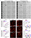
Fig. 2. NMII inhibition enhances neurite growth and branching.
Representative images of neurons cultured on CSPG (A) or laminin (B) 24, 48, and 72 h post incubation with DMSO, 25 μM blebbistatin, or 10 μM NMIIi2. Scale bar: 100 μm. Quantification of neurite length (mm/mm2) on CSPG (C) or laminin (D) substrates. Experiments performed in biological triplicates. Spot counts: 13 DMSO, 13 NMIIi2, and 12 blebbistatin on CSPG (C), and 12 DMSO, 12 NMIIi2, and 11 blebbistatin on laminin (D). Neurite length normalized to DMSO at 0 h. Statistical measurement two-way ANOVA with Tukey’s post hoc test, SEM error bars. Sample number and p-values as follows: CSPG - At 0 h: NMIIi2 (n = 13), Blebbistatin (n = 12). At 24 h: NMIIi2 (n = 13; p = < 0.0001), Blebbistatin (n = 12; p = 0.002). At 48 h: NMIIi2 (n = 13; p = <0.0001), Blebbistatin (n = 12; p = 0.003). At 72 h: NMIIi2 (n = 13; p = <0.0001), Blebbistatin (n = 12; p = 0.008). Laminin - At 0 h: NMIIi2 (n = 12), Blebbistatin (n = 11). At 24 h: NMIIi2 (n = 12; p = <0.0001), Blebbistatin (n = 11; p = 0.0009). At 48 h: NMIIi2 (n = 12; p = <0.0001), Blebbistatin (n = 11; p = 0.003). At 72 h: NMIIi2 (n = 12; p = 0.0006), Blebbistatin (n = 11; p = 0.01). Wells stained with an antibody against TUBB3 in CSPG (E) or laminin (F) after incubation with DMSO, 25 μM blebbistatin, or 10 μM NMIIi2. Scale bar: 75 μm. The arrowhead indicates branch points (yellow). Experiments conducted in biological triplicates. Sample number: 15 DMSO, 15 NMIIi2, and 12 blebbistatin on CSPG (G), and 14 DMSO, 14 NMIIi2, and 12 blebbistatin on laminin (H). Neurite length and branch points were quantified per neuronal count per field and normalized to DMSO for each condition. Stats: nonparametric Kruskal–Wallis test with Dunn’s multiple comparison test or two-sided Welch’s test with Dunnett’s multiple comparison test. SEM error bars are shown. P-values are indicated in the graph. Data for Fig. (C, D, G, and H) are available in the Source Data file.
Fig. 3
Fig. 3. NMII inhibition promotes regeneration after axonal injury.
A Image of a day 7 spot culture of human motor neurons with neurites projecting unidirectionally from cell bodies in the center. A laser cut injury was applied ~ 200 μm from the edge of the cell body cluster (proximal site injury) or ~ 700 µm from the cell body cluster, near the axon terminals (distal site injury). Injury size: 200–300 μm. Axon density was measured using a high-throughput SNT script that crops the injury site, adjusts the threshold, and binarizes the raw images. Images of regenerating neurons after axonal injury at proximal (B) or distal (C) sites. Scale bar: 100 μm. Quantification of axon density after a proximal (D) or distal injury (E) with DMSO or 10μM NMIIi2. Experiments were in triplicates. Data from ≥ 2 fields per spot were quantified and normalized to DMSO. All experiments conducted in triplicate, stats: two-way ANOVA with Tukey’s post hoc test, SEM error bars. Spot counts and p-values as follows: Proximal injury (DMSO) – 0 h (n = 12), 4 h (n = 12), 8 h (n = 11), 12 h (n = 12), 16 h (n = 12), 20 h (n = 12), and 24 h (n = 11), and (NMIIi2) – 0 h (n = 13, p = 0.98), 4 h (n = 13, p = 0.4), 8 h (n = 12, p = 0.3), 12 h (n = 12, p = 0.04), 16 h (n = 13, p = 0.006), 20 h (n = 13, p = 0.006), and 24 h (n = 12, p = < 0.0001); distal injury (DMSO) – 0 h (n = 15), 4 h (n = 15), 8 h (n = 14), 12 h (n = 15), 16 h (n = 15), 20 h (n = 15), and 24 h (n = 13), and (NMIIi2) − 0 h (n = 16, p = 0.99), 4 h (n = 16, p = 0.3), 8 h (n = 15, p = 0.1), 12 h (n = 15, p = 0.01), 16 h (n = 15, p = 0.01), 20 h (n = 16, p = 0.0008), and 24 h (n = 14, p = < 0.0001). Data for Fig. (D, and E) are available in the Source Data file.
Fig. 4
Fig. 4. NMII inhibition of injured axons increases neurite length and branching.
A Schematic of local NMIIi2 incubation of motor neuron cell bodies/proximal axons or distal axons in a microfluidic device and of the site of the axonal injury, by BioRender. Zeng, X. (2025) https://BioRender.com/i63w966. B Images of neuronal projections passing across microfluidic device microgroove before and after axotomy. Regenerating axons labeled by Sir-tub 24h-post injury (C) Representative images of regenerated axons incubated with DMSO, 10 μM NMIIi2 in either the axonal or cell body site for 24 h after a laser cut axonal injury. Images by confocal microscope, microtubules labeled with Sir-tub. Scale bar: 75 μm. D, E Quantification of total branch length and branch points. Data normalized to DMSO. Results were compiled from 21 microfluidic devices for the control group, 16 for NMIIi2 treatment of distal axons, and 13 for NMIIi2 treatment of cell bodies. Experiment conducted in four replicates. Statistical analysis: nonparametric Kruskal–Wallis test with Dunn’s multiple comparison test for (D) and two-sided Welch’s test with Dunnett’s multiple comparison test for (E), SEM error bars. P-values are indicated on the graph. Data for Fig. (D, E) are available in the Source Data file.
Fig. 5
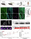
Fig. 5. NMIIi at sciatic nerve injury site accelerates motor function recovery in mice.
A NMIIi2 was delivered locally to sciatic nerve crush injury site packaged with a hydrogel scaffold in a silicone tube and motor behavior was assessed at different time points and lumbrical muscles dissected from mouse paw of day 14 post injury, schematic by BioRender. Zeng, X. (2025) https://BioRender.com/b18q366. B Representative images of lumbrical muscles day 14 post-injury from mice treated by local application of vehicle or NMIIi2 labeled with anti-neurofilament (NF), anti-synaptophysin (SYP) or α-bungarotoxin (BTX). C Co-localization of pre/post-synaptic markers per muscle in mice with intact sciatic nerve (non-injury) and 14 days post-nerve injury. Number of presynaptic markers that overlap with the postsynaptic marker, α-bungarotoxin normalized to post-synapse (α-bungarotoxin) number. Stats: nonparametric Kruskal–Wallis test with Dunn’s multiple comparison test, SEM error bars. P-values indicated on the graph. Data collected from 13 male mice/group. D Representative mouse paw luminance signals on day 14 post-injury. Paw on non-injured side (cyan), injured side (magenta). E Density histogram of right and left paw surface contact on day 14 post-injury in response to local administration of vehicle or NMIIi2. F Quantification of luminance ratio of left/right paws post sciatic nerve crush. Stats: two-way ANOVA with Tukey’s post hoc test, SEM error bars, 8 male mice per condition. G Two-sided Pearson correlation between the paw luminance ratio and percentage of pre/post-synaptic colocalization from the same mouse shown in panel (C). Data collected from 16 male mice per group. Data for Fig. (C, F, and G) are available in the Source Data file.
Fig. 6
Fig. 6. NMIIi2 applied at the sciatic nerve injury site promotes sensory axon regeneration and functional recovery.
A and B Representative images of mouse sciatic nerves day 3 post-injury treated with local application of either vehicle or NMIIi2 and labeled with anti-CGRP (green), anti-stathmin (magenta) (A), and anti-neurofilament (green) (B), white dashed lines indicate the boundary of the crush injury site. C Quantification of CGRP and stathmin intensity from regenerated axons. Data collected from 3 mice per group and normalized to vehicle condition. Statistics: unpaired two-tailed Student’s t-test, SEM error bars. D Score of pinprick tests following local application of vehicle or NMIIi2 at baseline and day 4, 7, and 14 post-injury. Statistics: two-way ANOVA with Tukey’s post hoc test, SEM error bars, 15 mice per condition. E Two-sided Pearson correlation analysis of the paw luminance ratio (Injured/Non-injured) and pinprick behavior scores on day 14 post-injury. Data collected from 10 male mice per group. Data for Fig. C-E are available in the Source Data file.

References

    1. Jain, N. B. et al. Traumatic spinal cord injury in the United States, 1993-2012. JAMA313, 2236–2243 (2015). - PMC - PubMed
    1. Gordon, T., Tyreman, N. & Raji, M. A. The basis for diminished functional recovery after delayed peripheral nerve repair. J. Neurosci.31, 5325–5334 (2011). - PMC - PubMed
    1. Grinsell, D. & Keating, C. P. Peripheral nerve reconstruction after injury: a review of clinical and experimental therapies. Biomed. Res Int.2014, 698256 (2014). - PMC - PubMed
    1. Lundborg, G. A 25-year perspective of peripheral nerve surgery: evolving neuroscientific concepts and clinical significance. J. Hand Surg. Am.25, 391–414 (2000). - PubMed
    1. Novak, C. B., Anastakis, D. J., Beaton, D. E., Mackinnon, S. E. & Katz, J. Biomedical and psychosocial factors associated with disability after peripheral nerve injury. J. Bone Jt. Surg. Am.93, 929–936 (2011). - PubMed

MeSH terms