. 2025 Mar 26;16(1):2949.
doi: 10.1038/s41467-025-58077-x.

Evidence supporting a catalytic pentad mechanism for the proteasome and other N-terminal nucleophile enzymes

Affiliations

Evidence supporting a catalytic pentad mechanism for the proteasome and other N-terminal nucleophile enzymes

Darlene Fung et al. Nat Commun. .

Abstract

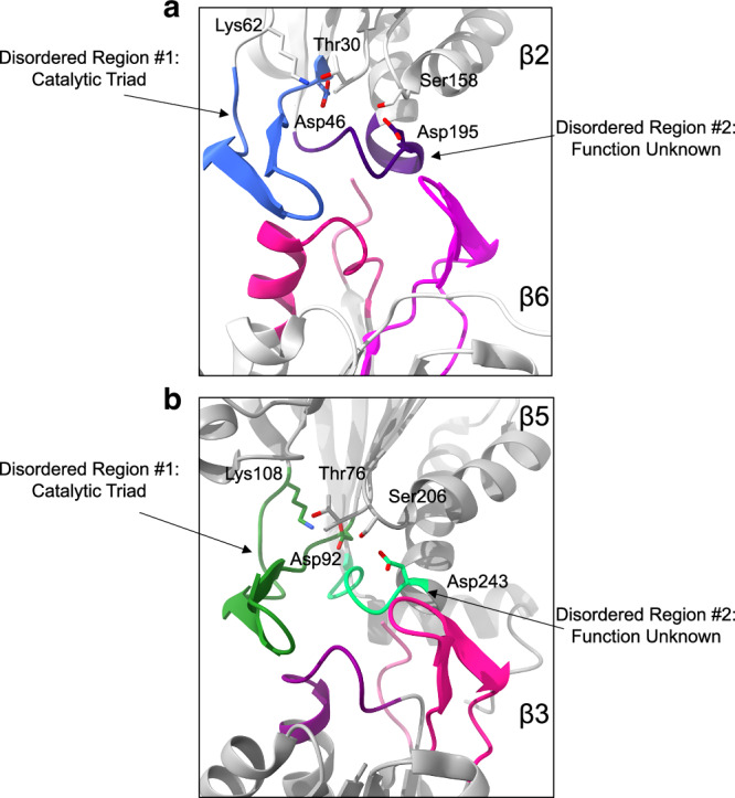
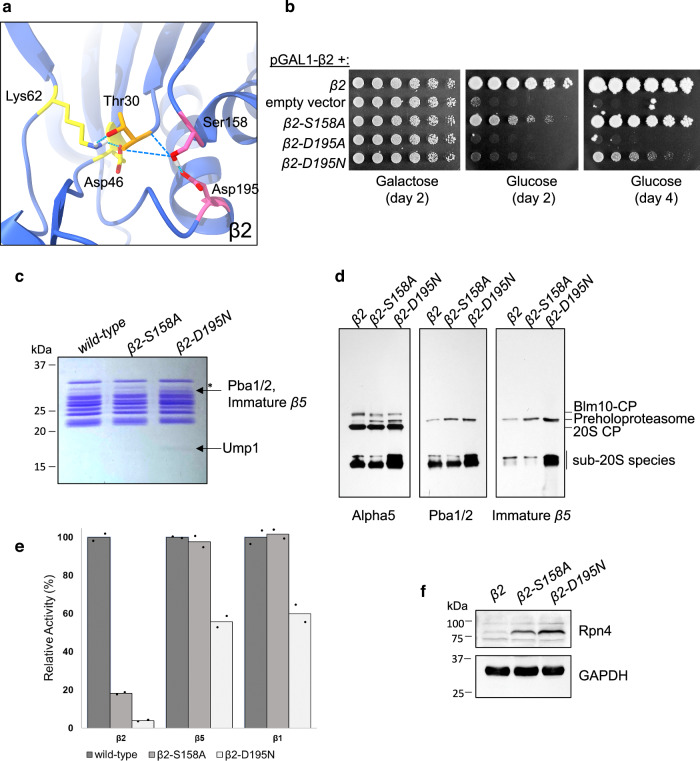
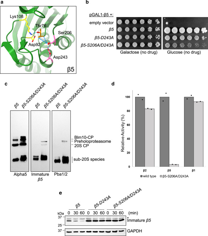
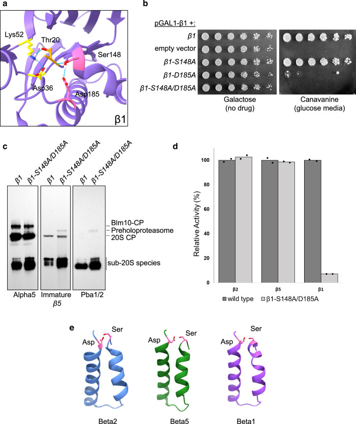
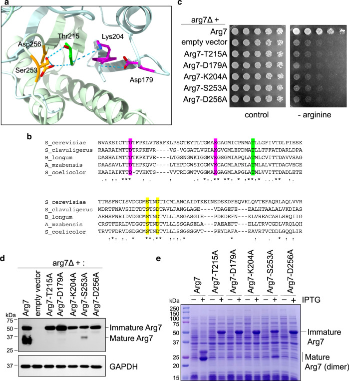
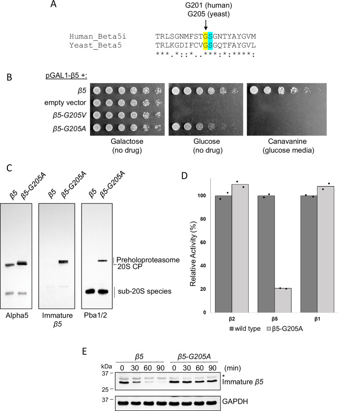
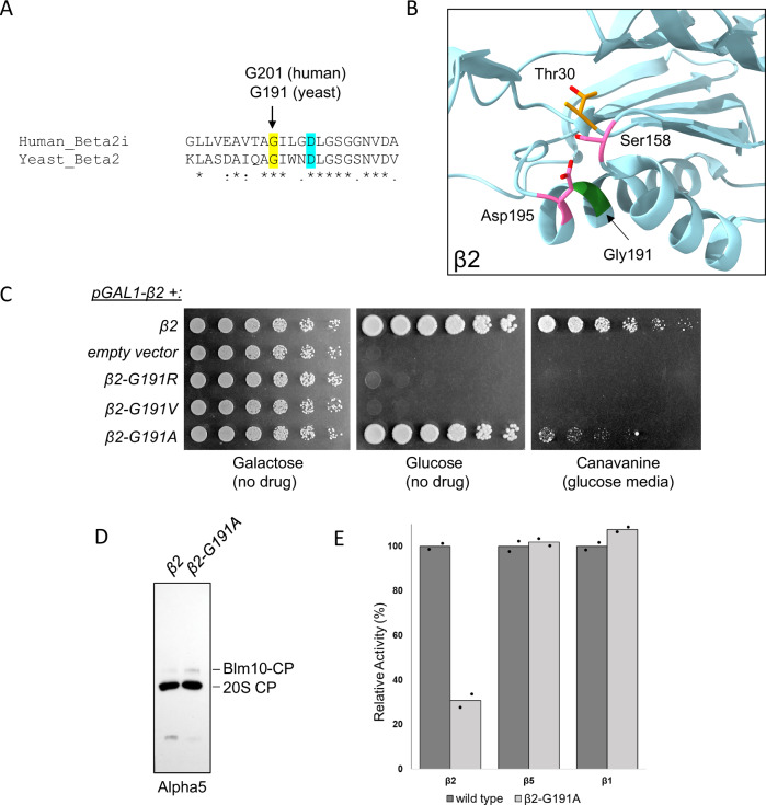
Proteases are defined by their nucleophile but require additional residues to regulate their active sites, most often arranged as catalytic triads that control the generation and resolution of acyl-enzyme intermediates. Threonine N-terminal nucleophiles represent a diverse family of proteases and transferases that possess two active site nucleophiles, the side chain hydroxyl and the free amino-terminus, and require autocatalytic cleavage of their N-terminal propeptides. Here we provide evidence that the proteasome, which mediates intracellular protein degradation and contains three different threonine protease subunits, utilizes a unique catalytic pentad mechanism. In addition to the previously defined lysine/aspartate pair which regulates threonine's side chain, a second serine/aspartate pair appears to regulate threonine's amino-terminus. The pentad is required for substrate proteolysis and assembly-coupled autocatalytic cleavage, the latter triggered by alignment of the full pentad upon fusion of two half-proteasome precursors. A similar pentad mechanism was required by the ornithine acetyltransferase Arg7, suggesting that this may be a general property of threonine N-terminal nucleophiles. Finally, we show that two patient-derived proteasome mutations compromise function of the serine/aspartate unit in yeast, suggesting that defective pentad function may underlie some human proteasomopathies.


Conflict of interest statement

Competing interests: The authors declare no competing interests.

Figures

Fig. 1
Fig. 1. Structural analysis of β-β-ring midline interactions.
Shown are the atomic models of wild-type CP (PDB: 5CZ4) with corresponding unresolved regions from the pre-15S assembly intermediate (PDB: 7LS6) shown in various colors. Panel (a) shows the β2/β6 interface. One unresolved loop in β2 appears to regulate the canonical threonine/lysine/aspartate catalytic triad, suggesting that stabilization of this loop may be a key feature of autocatalytic activation. However, β2 contains a separate unresolved loop whose function was unknown, and which contains a highly conserved aspartate residue with a nearby conserved serine residue, both in close proximity to the active site threonine. A similar arrangement was seen for the β5/β3 pair (b).
Fig. 2
Fig. 2. A catalytic pentad is required for autocatalytic activation and proteolysis by β2.
a Structural arrangement of the proposed pentad residues as seen in mature wild-type CP (PDB: 5CZ4). Lys62, Asp46, and Thr30 represent the canonical catalytic triad, while Ser158 and Asp195 represent additional residues proposed to complete the catalytic pentad. Dashed lines, hydrogen bonds. b Phenotypic analysis of the indicated mutants, expressed from low-copy centromeric plasmids harboring the endogenous promoter/terminator elements, in a strain where endogenous β2 is under the control of the pGAL1 promoter. In glucose-containing media, expression of the endogenous β2 locus is repressed and plasmid-derived β2 is the sole source of this protein. Plates were cultured at 30 °C for the indicated number of days. c Purified CP from wild-type and mutant strains, as analyzed using SDS-PAGE followed by Coomassie staining. Asterisk, a break-down product of α7. Note that Pba1, Pba2, and immature β5 co-migrate as a single band. d Analysis of wild-type and mutant CP by native gel electrophoresis followed by immunoblotting with antibodies against α5, Pba1/2, and immature (propeptide-bearing) β5. e Proteasome activity assays using active site-specific fluorescent substrate probes. Background fluorescence has been subtracted and relative activity has been normalized to untreated controls. Individual points represent biologic duplicates. f Levels of transcription factor Rpn4, which senses cellular proteasome capacity, in whole cell extracts from wild-type and mutant strains, as determined by SDS-PAGE and immunoblotting. GAPDH, loading control. Similar results were obtained in 5 (b), 4 (c, d) and 2 (f) independent experiments, respectively.
Fig. 3
Fig. 3. A catalytic pentad is required for autocatalytic activation and proteolysis by β5.
a Structural arrangement of the proposed pentad residues as seen in mature wild-type CP (PDB: 5CZ4). Lys108, Asp92, and Thr76 represent the canonical catalytic triad, while Ser206 and Asp243 represent additional residues proposed to complete the catalytic pentad. Dashed lines, hydrogen bonds. b Phenotypic analysis of the indicated mutants, expressed from low-copy centromeric plasmids harboring the endogenous promoter/terminator elements in a strain where endogenous β5 is under the control of the pGAL1 promoter. In glucose-containing media, expression of the endogenous β5 locus is repressed and plasmid-derived β5 is the sole source of this protein. Plates were cultured at 30 °C for 3 days. c Analysis of wild-type and mutant CP by native gel electrophoresis followed by immunoblotting with antibodies against α5, immature (propeptide-bearing) β5, and Pba1/2. d Proteasome activity assays using active site-specific fluorescent substrate probes. Background fluorescence has been subtracted and relative activity has been normalized to untreated controls. Individual points represent biologic duplicates. e Turnover of immature β5, as determined by cycloheximide chase analysis of whole cell extracts. Analysis was by SDS-PAGE followed by immunoblotting with an antibody that specifically recognizes immature β5. GAPDH, loading control. Asterisk, non-specific band. Similar results were obtained in 4 (b) and 2 independent experiments (c and e), respectively.
Fig. 4
Fig. 4. A catalytic pentad is required for autocatalytic activation and proteolysis by β1.
a Structural arrangement of the proposed pentad residues as seen in mature wild-type CP (PDB: 5CZ4). Lys52, Asp36, and Thr20 represent the canonical catalytic triad, while Ser148 and Asp185 represent additional residues proposed to complete the catalytic pentad. Dashed lines, hydrogen bonds. b Phenotypic analysis of the indicated mutants, expressed from low-copy centromeric plasmids harboring the endogenous promoter/terminator elements in a strain where endogenous β1 is under the control of the pGAL1 promoter. In glucose-containing media, expression of the endogenous β1 locus is repressed and plasmid-derived β1 is the sole source of this protein. Plates were cultured at 30 °C for 2 (no drug) and 7 days (canavanine, 1.5 μg/mL). c Analysis of wild-type and mutant CP by native gel electrophoresis followed by immunoblotting with antibodies against α5, immature (propeptide-bearing) β5, and Pba1/2. d Proteasome activity assays using active site-specific fluorescent substrate probes. Background fluorescence has been subtracted and relative activity has been normalized to untreated controls. Individual points represent biologic duplicates. e Structures of the helix-loop-helix motif responsible for the spatial proximity of the Ser/Asp pair within the catalytic pentad (PDB: 5CZ4). Similar results were obtained in 2 independent experiments (b, c).
Fig. 5
Fig. 5. A catalytic pentad is required for function and autocatalytic activity of the ornithine acetyltransferase Arg7.
a Structural arrangement of the proposed pentad residues as seen for Streptomyces clavuligerus ornithine acetyltransferase (PDB: 2VZK). For clarity, residue numbering is after yeast Arg7. Dashed lines represent hydrogen bonds. b Evolutionary conservation of the proposed catalytic pentad residues. c Phenotypic analysis of the indicated mutants, expressed from low-copy centromeric plasmids harboring the endogenous promoter/terminator elements in the arg7Δ mutant, which is strongly (although not fully) auxotrophic for arginine. Plates were cultured at 30 °C for 2 days. d Maturation of wild-type and mutant Arg7-3xHA proteins, as determined by analysis of whole cell extracts using SDS-PAGE followed by immunoblotting with anti-HA antibody. GAPDH, loading control. e Expression of recombinant wild-type and mutant Arg7 in bacteria, as analyzed by SDS-PAGE followed by Coomassie staining. Gene expression is induced by IPTG. The dimer seen for mature Arg7 reflects autocatalytic cleavage into two comparably sized polypeptides. Similar results were obtained in 2 (b) and 3 independent experiments (d, e), respectively.
Fig. 6
Fig. 6. Modeling of a patient-derived β5 mutation in yeast.
A Sequence alignment of human immunoproteasome β5i and yeast β5. The pentad serine residue is in cyan. B Phenotypic analysis of the indicated mutants, expressed from low-copy centromeric plasmids harboring the endogenous promoter/terminator elements in a strain where endogenous β5 is under the control of the pGAL1 promoter. In glucose-containing media, expression of the endogenous β5 locus is repressed and plasmid-derived β5 is the sole source of this protein. Plates were cultured at 30 °C for 2 (galactose), 1 (glucose), and 5 (canavanine 1.5 μg/mL) days. C Analysis of wild-type and mutant CP by native gel electrophoresis followed by immunoblotting with antibodies against α5, immature (propeptide-bearing) β5, and Pba1/2. D Proteasome activity assays using active site-specific fluorescent substrate probes. Background fluorescence has been subtracted and relative activity has been normalized to untreated controls. Individual points represent biologic duplicates. E Turnover of immature β5, as determined by cycloheximide chase analysis of whole extracts. Analysis was by SDS-PAGE followed by immunoblotting with the antibody that specifically recognizes immature β5. GAPDH, loading control. Asterisk, non-specific band. Similar results were obtained in 3 (B) and 2 independent experiments (C and E), respectively.
Fig. 7
Fig. 7. Modeling of a patient-derived β2 mutation in yeast.
A Sequence alignment of human immunoproteasome β2i and yeast β2. The pentad aspartate residue is in cyan. B Position of the corresponding G191 residue in yeast CP. An amino acid side chain at this position would be predicted to disrupt the Ser/Asp pair of the catalytic pentad. C Phenotypic analysis of the indicated mutants, expressed from low-copy centromeric plasmids harboring the endogenous promoter/terminator elements in a strain where endogenous β2 is under the control of the pGAL1 promoter. In glucose-containing media, expression of the endogenous β2 locus is repressed and plasmid-derived β2 is the sole source of this protein. Plates were cultured at 30 °C for 2 (galactose and glucose) and 7 (canavanine, 2 μg/mL) days. D Analysis of wild-type and mutant CP by native gel electrophoresis followed by immunoblotting with α5 antibody. E Proteasome activity assays using active site-specific fluorescent substrate probes. Background fluorescence has been subtracted and relative activity has been normalized to untreated controls. Individual points represent biologic duplicates. Similar results were obtained in 3 (C) and 2 independent experiments (D), respectively.

Similar articles

References

    1. Brannigan, J. A. et al. A protein catalytic framework with an N-terminal nucleophile is capable of self-activation. Nature378, 416–419 (1995). - PubMed
    1. Seemuller, E. et al. Proteasome from thermoplasma acidophilum: a threonine protease. Science268, 579–582 (1995). - PubMed
    1. Seemuller, E., Lupas, A. & Baumeister, W. Autocatalytic processing of the 20S proteasome. Nature382, 468–471 (1996). - PubMed
    1. Groll, M. et al. Structure of 20S proteasome from yeast at 2.4 A resolution. Nature386, 463–471 (1997). - PubMed
    1. Buller, A. R. & Townsend, C. A. Intrinsic evolutionary constraints on protease structure, enzyme acylation, and the identity of the catalytic triad. Proc. Natl Acad. Sci. USA110, 653–661 (2013). - PMC - PubMed

MeSH terms

LinkOut - more resources