. 2025 May;641(8064):1029-1036.
doi: 10.1038/s41586-025-08799-1. Epub 2025 Mar 26.

Gene-modified pig-to-human liver xenotransplantation

Affiliations

Gene-modified pig-to-human liver xenotransplantation

Kai-Shan Tao et al. Nature. 2025 May.

Abstract

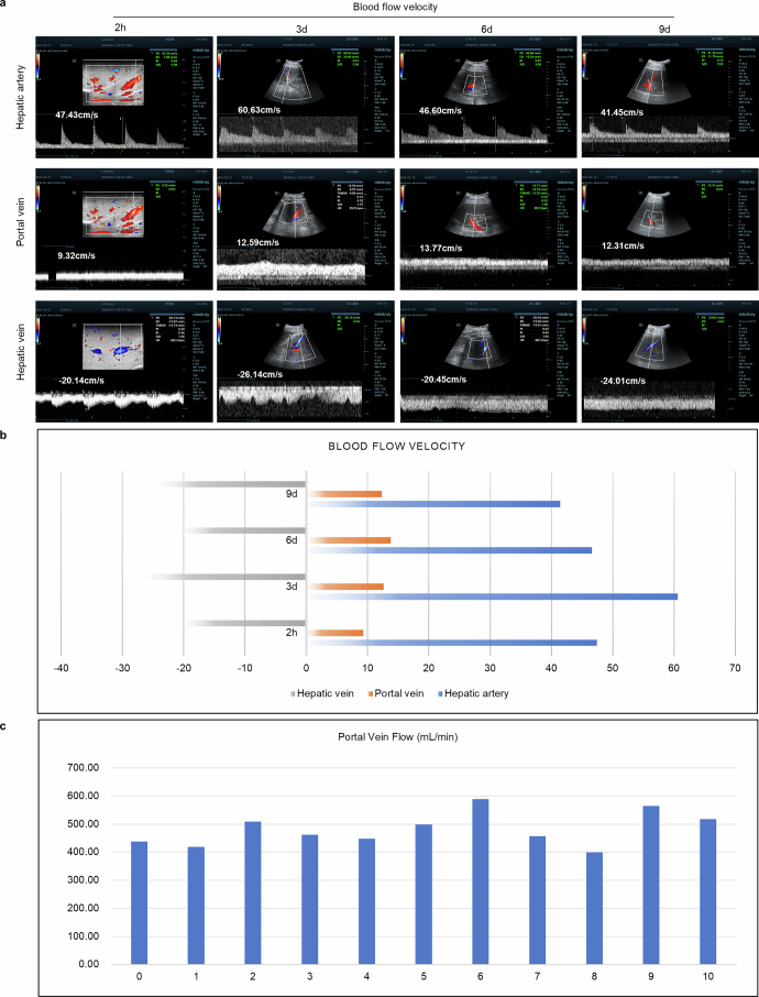
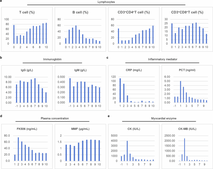
The shortage of donors is a major challenge for transplantation; however, organs from genetically modified pigs can serve as ideal supplements1,2. Until now, porcine hearts and kidneys have been successively transplanted into humans3-7. In this study, heterotopic auxiliary transplantation was used to donate a six-gene-edited pig liver to a brain-dead recipient. The graft function, haemodynamics, and immune and inflammatory responses of the recipient were monitored over the subsequent 10 days. Two hours after portal vein reperfusion of the xenograft, goldish bile was produced, increasing to 66.5 ml by postoperative day 10. Porcine liver-derived albumin also increased after surgery. Alanine aminotransferase levels remained in the normal range, while aspartate aminotransferase levels increased on postoperative day 1 and then rapidly declined. Blood flow velocity in the porcine hepatic artery and portal and hepatic veins remained at an acceptable level. Although platelet numbers decreased early after surgery, they ultimately returned to normal levels. Histological analyses showed that the porcine liver regenerated capably with no signs of rejection. T cell activity was inhibited by anti-thymocyte globulin administration, and B cell activation increased 3 days after surgery and was then inhibited by rituximab. There were no significant peri-operative changes in immunoglobulin G or immunoglobulin M levels. C-reactive protein and procalcitonin levels were initially elevated and then quickly declined. The xenograft remained functional until study completion.


Conflict of interest statement

Competing interests: The authors declare no competing interests.

Figures

Fig. 1
Fig. 1. Gene editing and pathogenic surveillance of the donor pig.
a, Donor pig. b, Flow cytometry result showing the numbers of GGTA1-, β4GalNT2-, Neu5Gc-, CD46- and CD55-positive cells in the donor pig and a wild-type (WT) pig. For gating strategies, see Supplementary Fig. 2. c, Western blotting result showing the levels of human CD46 and CD55 protein in the liver tissues of the donor and a wild-type pig. Glyceraldehyde 3-phosphate dehydrogenase (GAPDH) is used as the control. For blot source data, see Supplementary Fig. 1. d, IHC staining result showing the levels of human CD46, CD55 and thrombomodulin protein in the liver tissues of the donor and a wild-type pig. Positive signals are represented by black arrows. e, Flow cytometry result showing the ability of PBMCs, isolated from the donor and the wild-type pig, to bind human IgM and IgG, represented by median fluorescence intensity (MFI). For gating strategies, see Supplementary Fig. 3. f, Quantitative result of e. g, Relative (upper) and absolute (lower) quantification of real-time polymerase chain reaction (RT-PCR) showing the number of PERV copies in the PBMCs and liver tissues of the recipient and the donor pig. h, Relative quantification of RT-PCR showing PCMV quantity in the liver tissues of the recipient and the donor pig. bh contain one biological and technical repetition. Two independent experiments were carried out in c and d. Scale bar, 50 μm. CT, cycle threshold; ND, not detected. Source Data
Fig. 2
Fig. 2. Xenograft function.
a, Schematic depiction of the study, showing various time points of the investigation. b, Photographs of the bile secreted from the xenograft at different time points throughout the study. c,d, Amounts of bile (c) and porcine albumin (d) produced by the xenograft at different time points throughout the study. ek, Amounts of ALT (e), AST (f), total bilirubin (g), direct bilirubin (h), indirect bilirubin (i), γ-GGT (j) and alkaline phosphatase (k) in the recipient’s serum at different time points throughout the study. ln, Amounts of PLT (l), PT (m) and APTT (n) of the recipient at different time points throughout the study. cn contain one biological and technical repetition. DBil, direct bilirubin; IDBil, indirect bilirubin; IU, international unit; TBil, total bilirubin. Source Data
Fig. 3
Fig. 3. Histology of the xenograft and the original human liver.
a, Representative images of haematoxylin and eosin (H&E) staining of liver tissues obtained from the xenograft and the recipient at different time points. b, Representative images of IHC staining of Ki67 and alpha smooth muscle actin (αSMA), immunofluorescence staining of CD31 and 4′,6-diamidino-2-phenylindole (DAPI) and SEM observation of the hepatic sinusoid of the xenograft at different time points. Positive signals are shown with black or white arrows. c, Representative TEM images of the porcine hepatocytes at different time points. ac contain one biological and technical repetition. The experiment was carried out once in a, whereas two independent experiments were carried out in b and c. Scale bars, 1 μm (b (right), c (bottom)), 4 μm (c (top)), 25 μm (b (left)), 100 μm (a (bottom), b (middle)), 200 μm (a, top).
Fig. 4
Fig. 4. Immune and inflammatory monitoring of the recipient.
a, Representative images of IHC staining of C3d, C4d and C5b-9 on the liver sections obtained from the xenograft at different time points of the study. b, Representative images of IHC staining of IgM and IgG on the liver sections obtained from the xenograft at different time points of the study. cf, Amounts of interleukin-6 (IL-6) (c), TNF (d), IFNα (e) and IFNγ (f) in the recipient’s serum at different time points throughout the study. a and b contain one biological and technical repetition. For a, two independent experiments were carried out, whereas for b, the experiment was carried out once. Scale bars, 50 μm. Source Data
Extended Data Fig. 1
Extended Data Fig. 1. Pathogenic surveillance of the donor pig.
a, The PCR combined with gel electrophoresis detecting the presence of PCMV in the PBMCs of the recipient and the xenograft. b, The PCR combined with gel electrophoresis detecting the presence of PERV as well as microchimerism in the PBMCs of the recipient and the xenograft. PERV-ABC, the sequence shared by PERV-A, PERV-B and PERV-C. PERV-C, the sequence specifically belongs to PERV-C. RPP30 was used as the control gene. NTC, no template control. For gel source data, see Supplementary Fig. 1. Extended Data Fig. 1a,b contains 1 biological and technical repetition, and 3 independent experiments were carried out.
Extended Data Fig. 2
Extended Data Fig. 2. The heterotopic auxiliary liver xenotransplantation procedure.
a, The diameters of the portal vein (PV) and the inferior vena cava (IVC) of the recipient and the donor pig detected by vessel segmentation and ultrasound. b, Photos of the donor liver at different time points throughout the study. c, Photos of the vascular anastomosis between the recipient and the donor liver during surgery. d, A schematic depiction of the surgery. ①, ② and ③ represent the vascular anastomosis captioned in c. Source Data
Extended Data Fig. 3
Extended Data Fig. 3. Other surgery details.
a, b, Images of vessel segmentation in the donor pig. c, A photo of the recipient’s abdomen, showing the surgical incision, abdominal drains, and bile drain. d, A photo of the reconstruction of the recipient’s IVC by artificial vessel once the porcine liver was removed. e, The ultrasound-detected blood flow velocity of IVC after artificial vessel reconstruction.
Extended Data Fig. 4
Extended Data Fig. 4. Hemodynamic monitoring of the xenograft.
a, The ultrasound-detected blood flow velocity of the hepatic artery, the portal vein, and the hepatic vein of the xenograft at different time points of the study. The exact speed of blood flow is labelled correspondingly. b, The quantitative result of a. c, The ultrasound-detected portal vein flow (PVF) of the xenograft at different time points of the study. Source Data
Extended Data Fig. 5
Extended Data Fig. 5. Bleeding and coagulation monitoring of the recipient.
a, The indicators of coagulation function of the recipient at different time points of the study. b, The serum levels of coagulation factors of the recipient at different time points of the study. Extended Data Fig. 5a,b contains 1 biological and technical repetition. Source Data
Extended Data Fig. 6
Extended Data Fig. 6. Immune strategy of the recipient.
The immunosuppression strategy adopted in this study.
Extended Data Fig. 7
Extended Data Fig. 7. Immune monitoring of the recipient.
a, The levels of total T, B, CD3+CD4+ T and CD3+CD8+ T cells in the recipient’s blood at different time points of the study. b, The levels of IgM and IgG in the serum of the recipient at different time points of the study. c, The levels of C-reactive protein (CRP) and procalcitonin (PCT) in the serum of the recipient at different time points of the study. d, The plasma concentrations of FK506 and mycophenolate mofetil (MMF) of the recipient at different time points of the study. e, The serum levels of myocardial enzymes of the recipient at different time points of the study. Extended Data Fig. 7a–e contains 1 biological and technical repetition. Source Data

References

    1. Pan, D. et al. Progress in multiple genetically modified minipigs for xenotransplantation in China. Xenotransplantation26, e12492 (2019). - PubMed
    1. Cooper, D. K., Ekser, B., Ramsoondar, J., Phelps, C. & Ayares, D. The role of genetically engineered pigs in xenotransplantation research. J. Pathol.238, 288–299 (2016). - PMC - PubMed
    1. Griffith, B. P. et al. Genetically modified porcine-to-human cardiac xenotransplantation. N. Engl. J. Med.387, 35–44 (2022). - PMC - PubMed
    1. Montgomery, R. A. et al. Results of two cases of pig-to-human kidney xenotransplantation. N. Engl. J. Med.386, 1889–1898 (2022). - PubMed
    1. Loupy, A. et al. Immune response after pig-to-human kidney xenotransplantation: a multimodal phenotyping study. Lancet402, 1158–1169 (2023). - PubMed

MeSH terms