. 2025 Jun;642(8066):165-173.
doi: 10.1038/s41586-025-08797-3. Epub 2025 Mar 26.

The P-loop NTPase RUVBL2 is a conserved clock component across eukaryotes

Affiliations

The P-loop NTPase RUVBL2 is a conserved clock component across eukaryotes

Meimei Liao et al. Nature. 2025 Jun.

Erratum in

Abstract

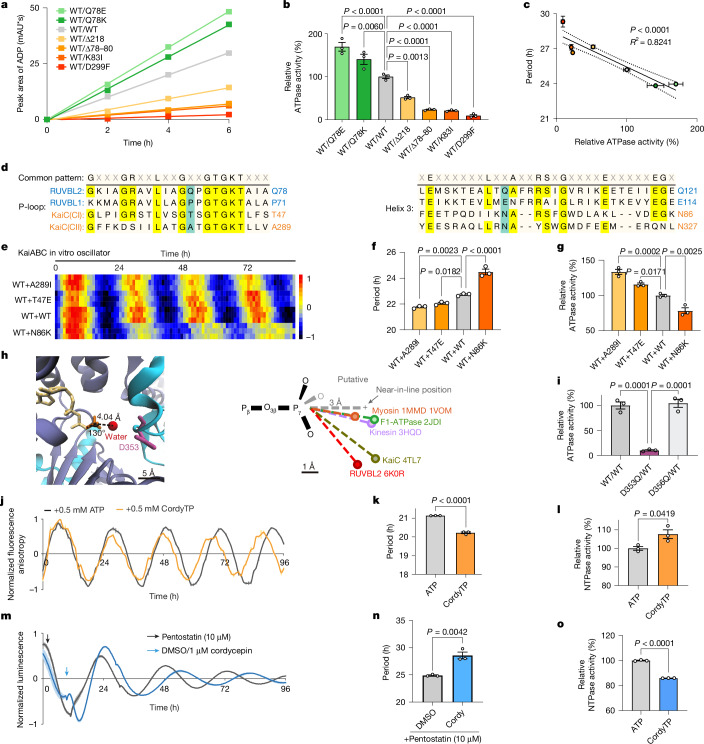
The eukaryotic circadian clock keeps time by using a transcription-translation feedback loop, which exhibits an architecture that is conserved across a diverse range of organisms, including fungi, plants and animals1. Despite their mechanistic similarity, the molecular components of these clocks indicate a lack of common ancestry2. Our study reveals that RUVBL2, which is a P-loop NTPase enzyme previously shown to affect circadian phase and amplitude as part of mammalian clock super-complexes, influences the circadian period through its remarkably slow ATPase activity, resembling the well-characterized KaiC-based clock in cyanobacteria. A screen of RUVBL2 variants identified arrhythmic, short-period and long-period mutants that altered circadian locomotor activity rhythms following delivery by adeno-associated virus to the murine suprachiasmatic nucleus. Enzymatic assays showed that wild-type RUVBL2 hydrolyses only around 13 ATP molecules a day, a vastly reduced turnover compared with typical ATPases. Notably, physical interactions between RUVBL2 orthologues and core clock proteins in humans, Drosophila and the fungus Neurospora, along with consistent circadian phenotypes of RUVBL2-mutant orthologues across species, reinforce their clock-related function in eukaryotes. Thus, as well as establishing RUVBL2 as a common core component in eukaryotic clocks, our study supports the idea that slow ATPase activity, initially discovered in cyanobacteria, is a shared feature of eukaryotic clocks.


Conflict of interest statement

Competing interests: E.E.Z. and D.J. have applied for a patent associated with this study (WIPO publication WO2018133835A1, Nucleoside analog regulating mammalian circadian rhythm).

Figures

Fig. 1
Fig. 1. A genetic screen unveils circadian mutant variants of RUVBL2 in U2OS cells.
a, The genetic screen procedure for circadian phenotypes on a specific gene. b, Schematic representation of single guide RNA (sgRNA) site design on coding sequences of PER2, ACTB and RUVBL2. c, Distribution of circadian parameters in mutants, with upper and lower panels representing period and amplitude distributions, respectively; mutRUVBL2 (n = 822), mutPER2 (n = 210), mutACTB (n = 298). The cut-off for the period is determined using log2(period/mean of mutACTB (25.75 h)). Mutants with a period of less than 24.03 h (log2 value < −0.1) are classified as short hits (green dots), those greater than 27.59 h (log2 value > 0.1) as long hits (orange) and those exceeding 33.98 h (log2 value > 0.4) as arrhythmic (blue). Amplitude is presented as log2(amplitude/mean of mutACTB), and the cut-off was −1.491 and +1.491 for low-amplitude hits (green) and high-amplitude hits (orange). d, Heatmap of Per2-dLuc expression of mutGenes ordered by phase. Each row represents one mutant; data of mutRUVBL2 were normalized between 4 (red) and −4 (blue). For mutRUVBL2, 822 mutants were divided into four groups, with 302 long-period mutants, 443 normal-period mutants, 22 short-period mutants and 55 arrhythmic mutants. e, Luminescence of three representative knock-in Per2-dLuc U2OS mutant cells: wild type (grey, shown in each graph for comparison), short period (green), long period (orange) and arrhythmic (AR, blue); n = 3 biologically independent cells. f, Period-length frequency distribution histogram of mutGenes. g, The proportions of period lengths. h, The distribution of 30 verified in-frame mutations in the RUVBL2 amino acid sequence. Residues for domain I (DI), domain II (DII) and domain III (DIII) are indicated, as well as catalytic motifs. All period data parameters were analysed using MetaCycle. Illustration in a created using BioRender (credit: Juan, C. https://BioRender.com/n10u764; 2024). Source Data
Fig. 2
Fig. 2. Mouse locomotor activities align with the observed period change or rhythmicity behaviours in the cellular assay.
a, Luminescence of representative knock-in Per2-dLuc U2OS mutants. The grey line represents RUVBL2WT/WT Per2-dLuc U2OS; n = 3 biologically independent cells. b, Locomotor activity of mice with introduced SCN-specific Ruvbl2 mutations. Top, the actogram of mice; bottom, the corresponding periodogram analysis. The grey line represents mice with GFP introduction in the SCN; n = 7, 8, 9 or 11 biologically independent mice for AAV–WT, K83, 78 K/78E/Δ120–121 and GEP/Δ78–80, respectively. LD indicates 12 h light:12 h dark; DD, always dark; grey shading indicates times of darkness. c, Left and middle: histograms of circadian periods in U2OS cells (left) and SCN-specific transgenic mice (middle). Right: simple linear regression analysis of periods. One-way analysis of variance (ANOVA) differential analysis using Šídák’s multiple comparisons test. d, Locomotor activity of SCN-specific Ruvbl2 knockout mice. Grey shading shows when the lights are off. The control and experimental group had 10 and 12 mice, respectively. e, Luminescence of PER2::LUC SCN with Ruvbl2 knockout in SCN. The right panel indicates amplitude analysis in PER2::LUC SCN recorded ex vivo. Sample sizes: n = 3 biologically independent mice for the control and n = 5 biologically independent mice for the knockout group. All period data parameters were analysed using LumiCycle (c). All bar graphs show mean ± s.e.m. Source Data
Fig. 3
Fig. 3. Two conserved motifs link ATP hydrolysis activity to circadian rhythm in the proteins RUVBL and KaiC.
a,b, Ultra-high-performance liquid chromatography (UHPLC) determination (a) and relative ATPase activity (b) of RUVBL1WT/RUVBL2WT and RUVBL1WT/RUVBL2MUT proteins. c, Correlation between the ATPase activity of human RUVBL1/2 and the circadian period length of knock-in U2OS cells. d, Primary sequence alignment of human RUVBL1/2 and cyanobacterial KaiC proteins. The sites related to ATPase activity in this study are labelled in blue. The image was created using Snapgene (v.4.3.6). e,f, Effects of the T47E, N86K or A289I mutants of KaiC proteins on fluorescence rhythms in the IVO system. The reaction was at standard condition but 25% of wild-type KaiC was replaced by mutant KaiC. Fluorescence anisotropy (e) is indicated by the colour scale, and period frequency is shown in f. g, Relative ATPase activity of mixing KaiC proteins. h, The position of the water molecule in the ATP-binding pocket in the human RUVBL1/RUVBL2 protein structure (6K0R). i, Relative ATPase activity of the RUVBL1MUT/RUVBL2WT and RUVBL1WT/RUVBL2WT proteins. j,k, Effects of CordyTP on the fluorescence rhythms in the IVO system, showing the fluorescence anisotropy (j) and period frequency (k). l, Relative ATPase activity of KaiC with an additional 0.5 mM ATP or CordyTP. m,n, Effects of cordycepin on the luminescence rhythm in Per2-dLuc U2OS cells, showing normalized luminescence (m) and period frequency (n). o, Relative ATPase activity of RUVBL1/RUVBL2 with an additional 0.5 mM ATP/CordyTP. Sample sizes: n = 3 biologically independent proteins for each group (a, b, c, g, i, l and o); n = 3 biologically independent cells for each group (c, m and n); n = 3 biologically independent samples for each group (e, f, j and k). All period data parameters were analysed using MetaCycle (f and k) and Lumicycle Analysis (n). Statistical tests: simple linear regression analysis (c); one-way ANOVA differential analysis with Šídák’s multiple comparisons test (b, f, g and i); two-tailed t-test differential analysis (k, l, n and o). All graphs show mean ± s.e.m. Credit: h was adapted from ref. , American Association for the Advancement of Science. Source Data
Fig. 4
Fig. 4. RUVBL2 regulates the circadian period in a CRY-independent manner.
a,b, Normalized luminescence expression in RUVBL2Q78E/WT (left), RUVBL2WT/WT (middle) and RUVBL2K83I/WT (right) knock-in U2OS cells with CRY1 (a) and CRY2 (b) knockdown from siRNA. The light and dark curves for each colour represent control and high-dose siRNA, respectively. c,d, Circadian periods of knock-in U2OS cells with low (#1) and high (#2) doses of CRY1 (c) and CRY2 (d) siRNA for knockdown; AR, arrhythmic. Periods were calculated by LumiCycle Analysis; n = 3 biologically independent cells. e,f, Knockdown efficiency (left two columns) and effects on clock gene expression (right nine columns) of CRY1 (e) and CRY2 (f) siRNA in unsynchronized U2OS cells; mRNA levels were analysed by quantitative PCR (qPCR), with ACTB as a control; n = 3 technical replicates, representing 3 independent experiments. In parallel experiments, bioluminescence rhythms were recorded and circadian phenotypes were confirmed. Statistical tests: all period data parameters were analysed using LumiCycle Analysis; two-tailed multiple unpaired t-test differential analysis with two-stage step-up (the Benjamini, Krieger and Yekutieli method) (c); two-way ANOVA differential analysis with Dunnett’s multiple comparisons test (d). All bar graphs show mean ± s.e.m. Source Data
Fig. 5
Fig. 5. A conserved function of RUVBL proteins in eukaryotic clocks.
a, Western blot analyses of co-immunoprecipitation (co-IP) of clock proteins and RUVBL2 homologues, which represents three independent experiments; antibodies for co-IP and western blot analyses are indicated. For gel data, see Supplementary Fig. 1a. IB, immunoblotting; IP, immunoprecipitation. b, Volcano plot of the proteins enriched by PER::GFP and IGG at zeitgeber time (ZT)16; n = 2 biologically independent experiments; see Methods for more details. c, Effects of CB-6644 in Bmal1-dLuc U2OS cells; n = 3 biologically independent cells for each dose. Left, heatmap of luminescence rhythms that were monitored in the presence of a dose concentration of CB-6644; luminescence intensity is indicated by the colour scale. Right, statistical analysis of the period length change. d, Stacked bar chart showing the rhythmicity of D. melanogaster with ReptinWT and ReptinK79E expression in tim-positive cells. The percentages of rhythmicity (R) and arrhythmicity (AR) are shown on the graph. e, Period length of rhythmic (R) Drosophila; n = 42, 32, 37 and 33 for UAS-WT, UAS-K79E, tim-GAL4;UAS-WT and tim-GAL4;UAS-K79E, respectively. f, Representations of double-plotted averaged actograms of flies measured for 5 days in 12 h:12 h light: dark (LD) and 11 days in constant dark (DD); grey shaded areas represent darkness. The data were analysed using ClockLab Analysis software. g, Relative ATPase activities of PontinWT/ReptinK79E and PontinWT/ReptinWT proteins; n = 3 biologically independent proteins for each group. h, Model of the origins of circadian oscillatory systems. The schematic shows the origins of each organism studied, stemming from the last universal common ancestor (LUCA). The period length is regulated by the activity of KaiC or RUVBL ATPase. Statistical tests: simple linear regression analysis (c); one-way ANOVA differential analysis with Šídák’s multiple comparisons test (e); two-tailed t-test differential analysis (g). All bar graphs show mean ± s.e.m. Illustrations in a and b adapted from BioRender (credit: Juan, C., https://BioRender.com/n10u764; 2024). Source Data
Extended Data Fig. 1
Extended Data Fig. 1. RUVBL2, with low ATPase activity, is conserved in eukaryotes.
a, Left panel: phylogenetic tree schematic illustrating the evolution of RUVBL proteins. Evolutionary analyses were conducted in MEGA11. Right panel: reference name for homologous genes and proteins. b, Protein sequence alignment of RUVBL proteins. c, A list of literature revealing the interactions between core clock proteins with RUVBL proteins. d, UHPLC determination of ATP hydrolysis activities of human RUVBL2 and cyanobacterial KaiC proteins. These activities are corresponding to consuming 12.6 (RUVBL2) and 12.9 (KaiC) ATP molecules per day per ATPase molecule, respectively. Two-tailed t-test differential analysis. n = 3 biologically independent proteins for each group. All bar graphs show mean ± s.e.m. Illustration in a created using BioRender (credit: Juan, C. https://BioRender.com/n10u764; 2024). Source Data
Extended Data Fig. 2
Extended Data Fig. 2. A genetic screen of RUVBL2 in U2OS cells.
a, Normalized luminescence expression in Per2-dLuc U2OS cells with or without stable Cas9 expression, n = 3 biologically independent cells. b, The effects of siPER2 and siACTB on Per2-dLuc U2OS cells. Data were reanalyzed from Zhang, E.E. et al. Cell. c, Compilation of mutation information for represented RUVBL2 and PER2 mutants in Fig. 1f. d, Compilation of in-frame (left) and frame-shift (right) mutation information for RUVBL2 mutants in secondary validation. e, The procedure of validation across entire screen. All period data parameters were analyzed from LumiCycle Analysis. All graphs show mean ± s.e.m. Source Data
Extended Data Fig. 3
Extended Data Fig. 3. RUVBL2 is essential for circadian rhythm of locomotor activity and can be stimulated by external cues.
a, Histograms of circadian periods of SCN-specific transgenic mice. n = 7, 8, 9, 11 biologically independent mice for AAV-WT, Δ151-152, Δ86-87/Δ350-352/Δ83 and GFP. b, Schematic illustration of generating the SCN-specific knockout mice. c, RUVBL2 expression in the SCN. d, Histogram of RUVBL2-positive cells in the SCN. n = 7 and 13 biologically independent mice for control and treat group, position where dense nuclei appeared was defined as 0 μm. e, Periodgram of SCN-specific Ruvbl2 knockout mice. n = 10 and 12 biologically independent mice for control and treat group. f, Left panel: histograms of circadian periods in transgenic mice. Right panel: actograms of mice and the corresponding periodogram. Gray line represents Ruvbl2WT introduction in the SCN. n = 9 mice per group. g, The mRNA expression levels of Ruvbl2 in liver and muscle. Three technical replicates from a mixed sample of 5 mice. h, Statistical analysis of Per2 and Ruvbl2 expression in mouse liver, and blood glucose, G6P as a positive control in fasting-refed experiments. Three technical replicates from a mixed sample of three mice for gene expression experiment. n = 9 (ZT12), 6 (ZT15), 3 (ZT17) biologically independent mice for blood glucose. i, Expression of RUVBL2 and c-Fos in the SCN, light pulse stimulation at CT15, data were analyzed from 5 mice per group. All period data parameters were analyzed from ClockLab Analysis. Two-way ANOVA differential analysis with Dunnett’s multiple comparisons test (d). One-way ANOVA differential analysis with Šídák’s multiple comparisons test (f). Two-tailed multiple unpaired t-test differential analysis with Two-stage step-up (Benjamini, Krieger, and Yekutieli) method (h). Two-tailed t-test differential analysis (i). All graphs show mean ± s.e.m. Illustration in a created using BioRender (credit: Juan, C. https://BioRender.com/n10u764; 2024). Source Data
Extended Data Fig. 4
Extended Data Fig. 4. ATP hydrolysis activity of RUVBL and KaiC proteins governs the clock period.
a, SDS-PAGE image of human RUVBL1/2 and cyanobacterial KaiC proteins. For gel source data, see Supplementary Fig. 1b. b, Alignment of the secondary structures of human RUVBL1/2 and cyanobacterial KaiC proteins. c, Eukaryotic RUVBL1/2 (in the HslU-ClpAB/CTD-LonAB-RuvB clade) and cyanobacterial KaiC (RecA homolog) genes belong to the P-loop NTPase superfamily (Figure is adapted from Erzberger et al Ann Rev Biophys Biomol Struct 2006). d, The structures of cordycepin triphosphate and adenosine triphosphate. e, Dose effects of cordycepin on the luminescence rhythms in Per2-dLuc U2OS cells in the presence of pentostatin, which prevents the rapid degradation of cordycepin in the human cells. n = 3 biologically independent cells. One-way ANOVA differential analysis with Šídák’s multiple comparisons test. All period data parameters were analyzed from Lumicycle Analysis. All bar graphs show mean ± s.e.m. Credit: c adapted with permission from ref. , Annual Reviews. Source Data
Extended Data Fig. 5
Extended Data Fig. 5. The circadian period of MAF cells were regulated by RUVBL2 in a CRY-independent manner.
a, The Ruvbl2Q78K/WT MAF cells were constructed by CRISPR-Cas9 method. b, c, Luminescence rhythm of Ruvbl2WT/WT and Ruvbl2Q78K/WT MAF cells. n = 3 biologically independent cells. d, Validation of CRY1 knock out efficiency and Cry2 knock out efficiency with western blot represents 3 independent experiments and Sanger sequencing results. Gel source data, see Supplementary Fig. 1c. e, Luminescence rhythm and period of WT MAF cells with Cry1 (left panel) or Cry2 (right panel) knock out. n = 3 biologically independent cells. f, Validation of CRY1 knock out efficiency (left panel) and Cry2 knock out efficiency (right panel) of Ruvbl2Q78K/WT MAF cells with western blot represents 3 independent experiments and Sanger sequencing. g, Luminescence rhythm and period of Ruvbl2Q78K/WT MAF cells with Cry1 or Cry2 knock out. n = 3 biologically independent cells. All period parameters were analyzed by LumiCycle Analysis. Two-tailed t-test differential analysis (c). One-way ANOVA differential analysis with Šídák’s multiple comparisons test (e, g). All graphs show mean ± s.e.m. Source Data
Extended Data Fig. 6
Extended Data Fig. 6. RUVBL2 interacts core clock proteins in eukaryotes.
Western blot analyses of co-immunoprecipitation (co-IP) of clock proteins from different species which represented 3 independent experiments, antibodies for co-IP and WB analyses are indicated. a, Co-IP by STREP. b, Co-IP by FLAG. For gel source data, see Supplementary Fig. 1d and e. Illustration in a created using BioRender (credit: Juan, C. https://BioRender.com/n10u764; 2024).
Extended Data Fig. 7
Extended Data Fig. 7. Inhibiting the ATPase activity of RUVBL2 with compound lengthens the circadian periods in multiple species.
a, The structure of CB-6644. b, The inhibitory effects of CB-6644 on the ATPase activity of human RUVBL1/2. c, Normalized luminescence rhythm of Bmal1-dLuc U2OS cells shows the period lengthening for 2 h in the presence of CB-6644 (3 μM), n = 3 biologically independent cells. d, Left panel: statistical analysis of the period length changes of Drosophila melanogaster. Right panel: represents of double-plotted averaged actogram of Drosophila melanogaster with CB-6644 or control treatment. n = 24 (DMSO), 22 (10 μM), 21 (25 μM), 23 (50 μM) biologically independent files. e, Left panel: statistical analysis of the period length of Neurospora crassa with CB-6644 treatment. Right panel: normalized luminescence rhythm of Neurospora crassa treated by CB-6644. n = 4 biologically independent fungi. f, Left panel: statistical analysis of the period length of Arabidopsis thaliana with CB-6644 treatment. Right panel: normalized luminescence rhythm of Arabidopsis thaliana treated by CB-6644, cordycepin, and adenosine. n = 3 (100 μM), 4 (DMSO/10/50 μM) biologically independent plants. The Drosophila melanogaster locomotor activity was analyzed by ClockLab Analysis. The period parameter of Neurospora crassa was analyzed from LumiCycle Analysis. The period parameter of Arabidopsis thaliana was analyzed from MetaCycle. One-way ANOVA differential analysis with Šídák’s multiple comparisons test (d, e, f). All bar graphs show mean ± s.e.m. Source Data
Extended Data Fig. 8
Extended Data Fig. 8. Reptin and Pontin knockdown in flies lengthens the circadian period or disrupts rhythmicity.
a, Quantification of percent rhythmicity for Pontin and Reptin RNAi knockdown flies. Black bar represents the % of rhythmic individuals and the grey bar represents the % of arrhythmic individuals in each genotype. b, Period of Drosophila lines with Pontin or Reptin RNAi expressed in all clock cells (crossed with tim-GAL4 driver flies), in Pigment Dispersing Factor (PDF) expressing brain neurons (crossed with pdf-GAL4 driver flies), or not expressed (crossed with w1118 flies). w1118 flies as control. n = 8, 16, 31, 14 for w1118, UAS-siRept#1;w1118, UAS-siRept#1;tim-GAL4 and UAS-siRept#1;pdf-GAL4. n = 11, 20, 31, 25 for w1118, UAS-siRept#2;w1118, UAS-siRept#2;tim-GAL4 and UAS-siRept#2;pdf-GAL4. n = 8, 11, 32, 9 for w1118, UAS-siPont#1;w1118, UAS-siPont#1;tim-GAL4 and UAS-siPont#1;pdf-GAL4. n = 11, 15, 15, 14 for w1118, UAS-siPont#2;w1118, UAS-siPont#2;tim-GAL4 and UAS-siPont#2;pdf-GAL4. In certain genotypes, the free-running period was calculated separately for the initial 4 days of DD (1-4 DD) and the last three days (5–7 DD) to detect arrhythmicity that was prominent after day four. The Drosophila melanogaster locomotor activity was analyzed by ClockLab Analysis. The data show mean ± s.e.m. Source Data
Extended Data Fig. 9
Extended Data Fig. 9. The rvb2 knock-in mutant Neurospora lost growth rhythm under constant-dark conditions.
a, The schematic illustration of introducing mutation on rvb2 (the RUVBL2 homolog) in Neurospora. b, Growth rhythm detection of rvb2S79I and rvb2WT knock-in mutant strains with race tube assays, and rvb2WT as a control. Two clones of rvb2S79I were presented. There are three conditions (upper: DD conditions, middle: light 12 h: dark 12 h conditions, and lower: 25 °C 12 h: 30 °C 12 h temperature cycles conditions) which were used to evaluate the impact of rvb2K79I on core clock of Neurospora. n is labeled next to it. n = 3 biologically independent fungi. c, The ATPase activity of RUVB1WT/RUVB2WT and RUVB1WT/ RUVB2S79I. n = 3 biologically independent proteins for each group. Two-tailed t-test differential analysis. All bar graphs show mean ± s.e.m. Source Data

References

    1. Zhang, E. E. & Kay, S. A. Clocks not winding down: unravelling circadian networks. Nat. Rev. Mol. Cell Biol.11, 764–776 (2010). - PubMed
    1. Rosbash, M. The implications of multiple circadian clock origins. PLoS Biol.7, e1000062 (2009). - PMC - PubMed
    1. Hardin, P. E., Hall, J. C. & Rosbash, M. Feedback of the Drosophila period gene product on circadian cycling of its messenger RNA levels. Nature343, 536–540 (1990). - PubMed
    1. Takahashi, J. S. Transcriptional architecture of the mammalian circadian clock. Nat. Rev. Genet.18, 164–179 (2017). - PMC - PubMed
    1. Johnson, C. H., Stewart, P. L. & Egli, M. The cyanobacterial circadian system: from biophysics to bioevolution. Annu. Rev. Biophys.40, 143–167 (2011). - PMC - PubMed

MeSH terms

Substances

LinkOut - more resources