. 2025 Mar 26;13(1):67.
doi: 10.1186/s40478-025-01977-2.

C9ORF72 poly-PR disrupts expression of ALS/FTD-implicated STMN2 through SRSF7

Affiliations

C9ORF72 poly-PR disrupts expression of ALS/FTD-implicated STMN2 through SRSF7

Karen S Wang et al. Acta Neuropathol Commun. .

Abstract

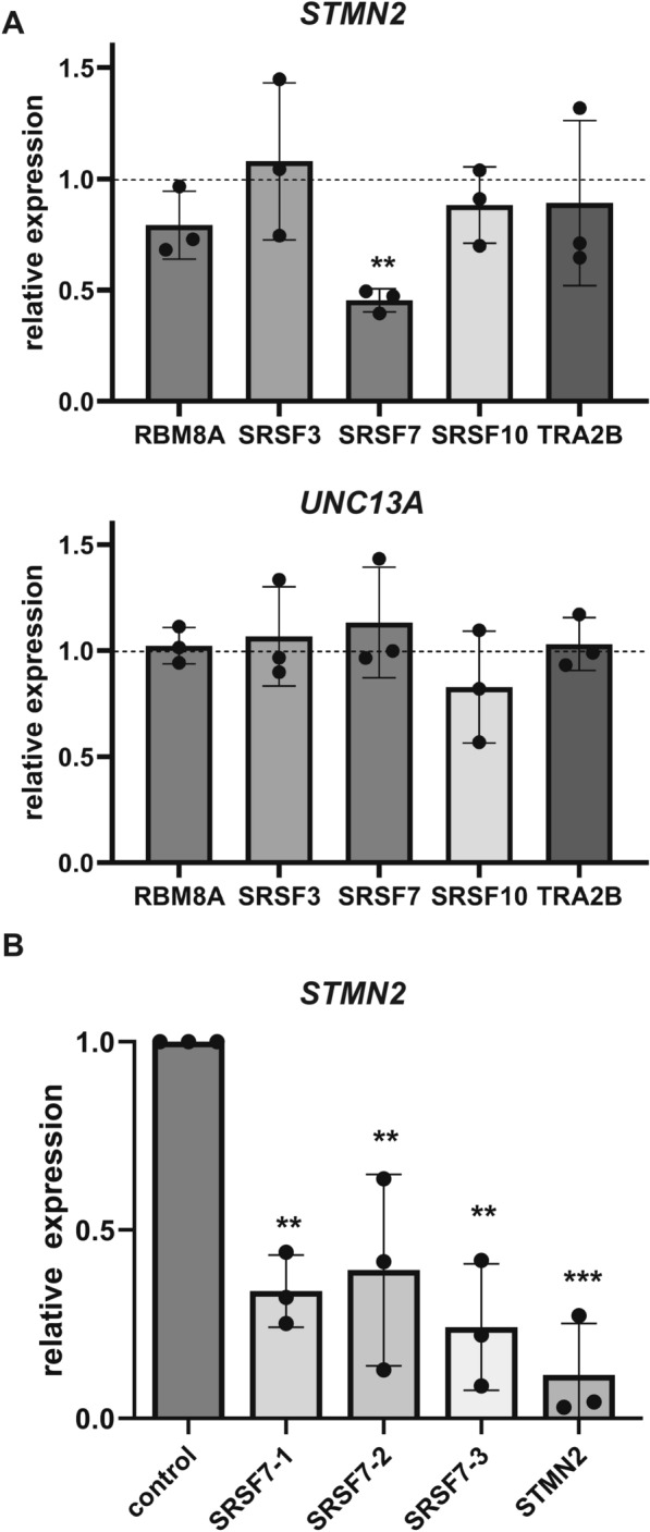
A hexanucleotide repeat expansion in C9ORF72 is the most common genetic cause of amyotrophic lateral sclerosis (ALS), frontotemporal dementia (FTD), and combined ALS/FTD. The repeat is transcribed in the sense and the antisense directions to produce several dipeptide repeat proteins (DPRs) that have toxic gain-of-function effects; however, the mechanisms by which DPRs lead to neural dysfunction remain unresolved. Here, we observed that poly-proline-arginine (poly-PR) was sufficient to inhibit axonal regeneration of human induced pluripotent stem cell (iPSC)-derived neurons. Global phospho-proteomics revealed that poly-PR selectively perturbs nuclear RNA binding proteins (RBPs). In neurons, we found that depletion of one of these RBPs, SRSF7 (serine/arginine-rich splicing factor 7), resulted in decreased abundance of STMN2 (stathmin-2), though not TDP-43. STMN2 supports axon maintenance and repair and has been recently implicated in the pathogenesis of ALS/FTD. We observed that depletion of SRSF7 impaired axonal regeneration, a phenotype that could be rescued by exogenous STMN2. We propose that antisense repeat-encoded poly-PR perturbs RBPs, particularly SRSF7, resulting in reduced STMN2 and axonal repair defects in neurons. Hence, we provide a potential link between DPRs gain-of-function effects and STMN2 loss-of-function phenotypes in neurodegeneration.


Conflict of interest statement

Declarations. Ethics approval and consent to participate: The usage of human induced pluripotent stem cells (iPSCs) was approved by the Human Gamete, Embryo and Stem Cell Research (GESCR) Committee at UCSF. Consent for publication: Not applicable. Competing interests: K.E. is a co-founder of Quralis and Enclear Therapies, and currently employed at BioMarin Pharmaceutical. D.A.M. receives sponsored research support from Genentech/Roche that is unrelated to this study. The remaining authors declare no competing interests.

Figures

Fig. 1
Fig. 1
Poly-PR decreases STMN2 expression in induced neurons and impairs axon regrowth. A Assessment of STMN2 and TARDBP relative expression via qPCR in human induced neurons transduced with lentivirus expressing GFP-(PR)50 or GFP only and then collected 6 days later. (** = p < 0.01, two-tailed t-test; n = 4 from two independent differentiations). B Schematic of axotomy assay in induced neurons in microfluidics chambers. C Representative fluorescent images of neurite regrowth of induced neurons transduced with lentivirus for GFP-(PR)50 or GFP only. D Quantification of neurite regrowth from three devices for each condition. (GFP-(PR)50 n = 147, GFP n = 108; **** p < 0.0001, two-tailed t-test)
Fig. 2
Fig. 2
Global phospho-proteomics reveals that poly-PR perturbs phospho-proteins involved in RNA processing. A Global phospho-proteomics mass spectrometry workflow of cells expressing GFP (control), GFP-(PR)50, GFP-(GR)50, collected from 4 independent transfections. B Volcano plot of the relative abundance of phospho-proteins from cells expressing GFP-(PR)50 versus GFP (control) and GFP-(GR)50 vs. GFP (control). C Gene ontology (GO) enrichment analysis of phospho-proteins increased in abundance in GFP-(PR)50, categorized by molecular function (MF), biological process (BP), or cellular component (CC). D Protein–protein interaction network of phospho-proteins increased in GFP-(PR)50. E Schematic of phosphorylation sites for the RNA processing factor SRSF7; RRM: RNA recognition motif, Zn Kn: Zinc knuckle domain, RS: Arginine/serine-rich domain. Changes in the relative abundance of selected SRSF7 phospho-peptides in GFP-(PR)50 (n = 3–4 per condition)
Fig. 3
Fig. 3
Knockdown of essential RNA processing factor SRSF7 decreases expression of STMN2 in induced neurons. A Representative RNA binding proteins identified from the global phospho-proteomics analysis were depleted in induced neurons, and expression of STMN2 and UNC13A were assessed via qPCR compared to non-targeting control (n = 3, ** = p < 0.01, unpaired two-tailed t-test). B STMN2 expression following transfection of induced neurons with non-targeting control siRNA or three individual siRNAs targeting SRSF7 across three independent differentiations. Pooled siRNAs against STMN2 served as a positive control. (** = p < 0.01, *** = p < 0.001, one-way ANOVA with Tukey’s correction for multiple comparisons)
Fig. 4
Fig. 4
Knockdown of essential SRSF7 reduces levels of STMN2, though not TDP-43, and impairs axon regrowth in induced neurons. A Representative immunoblot of STMN2, TDP-43, beta-tubulin IIIb (Tuj1) from induced neurons following depletion of SRSF7 or a non-targeting control. B Quantification of the levels of STMN2, TDP-43, and Tuj1 normalized to GAPDH, from three independent experiments (n = 7–8, *** = p < 0.001, two-tailed t-test). C Experimental workflow for axotomy assay. D Representative images of neurite regrowth following transfection of siRNAs targeting STMN2, SRSF7, or non-targeting control. E Axotomy assay with quantification of neurite regrowth from three separate devices for each condition (one-way ANOVA, **** = p < 0.0001). F Experimental workflow for rescue axotomy assay. G Axotomy assay of neurons transduced with lentivirus expressing STMN2-GFP compared to GFP control following siRNA transfection of non-targeting control or SRSF7 siRNA. Quantification of neurite regrowth, results from multiple separate devices for each condition (one-way ANOVA, **** = p < 0.0001)

Similar articles

References

    1. Brown RH, Al-Chalabi A (2017) Amyotrophic lateral sclerosis. N Engl J Med 377(2):162–172 - PubMed
    1. Taylor JP, Brown RH Jr, Cleveland DW (2016) Decoding ALS: from genes to mechanism. Nature 539(7628):197–206 - PMC - PubMed
    1. Renton AE et al (2011) A hexanucleotide repeat expansion in C9ORF72 is the cause of chromosome 9p21-linked ALS-FTD. Neuron 72(2):257–268 - PMC - PubMed
    1. DeJesus-Hernandez M et al (2011) Expanded GGGGCC hexanucleotide repeat in noncoding region of C9ORF72 causes chromosome 9p-linked FTD and ALS. Neuron 72(2):245–256 - PMC - PubMed
    1. Mori K et al (2013) The C9orf72 GGGGCC repeat is translated into aggregating dipeptide-repeat proteins in FTLD/ALS. Science 339(6125):1335–1338 - PubMed

Publication types

MeSH terms

Substances