. 2025 Mar 13;26(6):2583.
doi: 10.3390/ijms26062583.

Valproic Acid Improves Antisense-Mediated Exon-Skipping Efficacy in mdx Mice

Affiliations

Valproic Acid Improves Antisense-Mediated Exon-Skipping Efficacy in mdx Mice

Micky Phongsavanh et al. Int J Mol Sci. .

Abstract

Duchenne muscular dystrophy (DMD) is a severe genetic disorder characterized by the progressive degeneration of skeletal and cardiac muscles due to the absence of dystrophin. Exon-skipping therapy is among the most promising approaches for treating DMD, with several antisense oligonucleotides (ASO) already approved by the FDA; however, their limited efficacy highlights substantial potential for further improvement. In this study, we evaluate the potential of combining ASO with valproic acid (VPA) to enhance dystrophin expression and improve functional outcomes in a murine model of DMD. Our results indicate that the ASO+VPA treatment significantly increases dystrophin restoration across various muscle tissues, with particularly pronounced effects observed in cardiac muscle, where levels are nearly doubled compared to ASO monotherapy. Additionally, we demonstrate significant improvements in functional outcomes in treated mdx mice. Our findings suggest that the combined ASO+VPA therapy holds promise as an effective therapeutic approach to ameliorate muscle function in DMD, warranting further exploration of its mechanistic pathways and long-term benefits.

Keywords: RNA; antisense oligonucleotides; duchenne muscular dystrophy; exon skipping; histone deacetylase inhibitors; transcript imbalance; valproic acid.


Conflict of interest statement

L.G. is co-founder of SQY Therapeutics, which produces tricyclo-DNA oligomers. T.T. was an employee of SQY Therapeutics when performing this study. The remaining authors declare that the research was conducted in the absence of any commercial or financial relationships that could be construed as a potential conflict of interest.

Figures
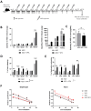
Figure 1
Figure 1
Long-term treatment with VPA increases exon-skipping efficacy. (A) Schematic representation of the treatment of mdx mice with ASO combined with valproic acid (VPA). (B) Effect of VPA on tcDNA-ASO distribution in various tissues. (C) Quantification of tcDNA-ASO in urines of treated mdx mice; urines were collected over a period of 24 h in metabolic cages. (D) Quantification of exon 23 skipping levels by TaqMan RT-qPCR in mdx mice treated with ASO or ASO+VPA. (E) Dystrophin restoration quantified by Western blot in mdx mice treated with ASO or ASO+VPA. (F) Effect of the combination VPA+ASO on Dmd transcript imbalance in diaphragm, and heart analyzed by TaqMan qPCR at different exon junctions. TA: tibialis anterior, GAS: gastrocnemius, QUAD: quadriceps, TRI: triceps and DIA: diaphragm. Results are expressed as the mean ± SEM; n = 6–8 mice per group, * p < 0.05, *** p < 0.001, **** p < 0.0001 and ns: non statistically significant analyzed by two-way ANOVA.
Figure 2
Figure 2
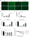
Functional recovery following the combined therapy ASO+VPA. (A) Detection of dystrophin protein (green staining) by immunostaining on transverse sections of muscle tissues (triceps and heart) from WT and mdx mice treated with saline, ASO, VPA or ASO+VPA. Nuclei are labelled with DAPI (blue staining). Scale bar, 100 µm. (B) Quantification of the dystrophin intensity staining in heart and triceps; n = 4 mice per group. (C) Myomesin-3 levels in serum of treated mice detected by Western blot; n = 5–7 mice per group. (D) Latency to fall in seconds in the inverted grid test; n = 6–9 mice per group. (E) Latency to fall in seconds in the wire test; n = 6–9 mice per group. (F) Maximal specific force in mg/g measured from the two tibialis anterior muscles of each mouse; n = 6–8 mice per group. (G) percentage of force drop following a series of 15 eccentric contractions measured on semi-isolated tibialis anterior muscles from treated mdx mice; n = 6–8 mice per group. Results are expressed as the mean ± SEM; * p < 0.05, *** p < 0.001, **** p < 0.0001 and ns: non statistically significant analyzed by two-way ANOVA.
Figure 3
Figure 3
Impact of long-term treatment with VPA in the brain. (A) Unconditioned fear response expressed as percentage of freezing (left panel), vertical activity (middle panel) or distance traveled (right panel) during a 5 min period of observation following a brief scruff restraint (15 s) (mean ± SEM; * p < 0.05; *** p < 0.005; **** p < 0.0001 analyzed by one-way ANOVA followed by Sidak post hoc tests). (B) Relative expression of Dmd transcript levels in mdx compared to WT levels quantified in cortex, hippocampus and cerebellum (CBL) at various exon–exon junctions along the Dmd gene (n = 8 per group) (* p < 0.05; ** p < 0.01 in cortex, *** p < 0.0001 in hippocampus and **** p < 0.0001 in cerebellum). (C) Relative expression of Dmd transcript levels in mdx mice treated with PBS (MDX), VPA, ASO, or the combination of ASO+VPA at various exon–exon junctions along the Dmd gene (n = 8 per group). (D) Quantification of the different dystrophins expression (Dp427, Dp140 and Dp71) in brain tissues (in cortex, hippocampus and cerebellum (CBL)) treated with VPA, ASO, or the combined therapy ASO+VPA by Western blot. The expression of each dystrophin is expressed as a percentage of levels detected in WT mice. Results are expressed as the mean ± SEM.
Figure 4
Figure 4
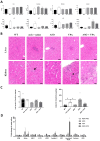
Evaluation of the safety profile of the combined ASO+VPA therapy. (A) Quantification of general toxicity biomarkers in the serum: creatinine, urea, albumin, aspartate aminotransferase (AST), alanine aminotransferase (ALT), alkaline phosphatase (ALP) and bilirubin; n = 7 mice per group, * p < 0.05, **, p < 0.01, and *** p < 0.001 compared to mdx saline, analyzed by the Kruskal–Wallis test. (B) Histopathological analysis from liver and kidney sections from WT and mdx mice treated with saline solution, ASO, VPA or ASO+VPA, stained with hematoxylin–eosin–saffron. In liver (upper panel), small foci of inflammatory cell infiltration were scattered in the hepatic parenchyma of all mice in every group. In kidney (lower panel), no lesions or only sporadic changes were observed in WT mice and mdx saline- and mdx-ASO-treated mice, whereas all VPA and ASO+VPA mice presented histopathological changes ranging from tubular degeneration/regeneration associated with various degree of proteinaceous casts (short arrow) and interstitial inflammation to tubular atrophy and loss and interstitial fibrosis. These changes included basophilic tubules, an early unspecific change that is a relatively common finding in aging mice and was present focally in one mdx saline mouse (arrow). Scale bar = 50 µm. (C) Quantification of total protein/creatinine (left panel) and albumin/creatinine (right panel) in urines of WT and mdx mice treated with saline solution, ASO, VPA or ASO+VPA. * p < 0.05 compared to mdx saline, analyzed by the Kruskal–Wallis test. (D) Quantification of kidney injury biomarkers in the urines of treated mice; n = 7 mice per group, two-way ANOVA analyses. Results are expressed as the mean ± SEM.

References

    1. Duan D., Goemans N., Takeda S., Mercuri E., Aartsma-Rus A. Duchenne Muscular Dystrophy. Nat. Rev. Dis. Primers. 2021;7:13. doi: 10.1038/s41572-021-00248-3. - DOI - PMC - PubMed
    1. Fortunato F., Rossi R., Falzarano M.S., Ferlini A. Innovative Therapeutic Approaches for Duchenne Muscular Dystrophy. J. Clin. Med. 2021;10:820. doi: 10.3390/jcm10040820. - DOI - PMC - PubMed
    1. Hammond S.M., Aartsma-Rus A., Alves S., Borgos S.E., Buijsen R.A.M., Collin R.W.J., Covello G., Denti M.A., Desviat L.R., Echevarría L., et al. Delivery of Oligonucleotide-Based Therapeutics: Challenges and Opportunities. EMBO Mol. Med. 2021;13:e13243. doi: 10.15252/emmm.202013243. - DOI - PMC - PubMed
    1. Roberts T.C., Wood M.J.A., Davies K.E. Therapeutic Approaches for Duchenne Muscular Dystrophy. Nat. Rev. Drug Discov. 2023;22:917–934. doi: 10.1038/s41573-023-00775-6. - DOI - PubMed
    1. Goyenvalle A., Griffith G., Babbs A., El Andaloussi S., Ezzat K., Avril A., Dugovic B., Chaussenot R., Ferry A., Voit T., et al. Functional Correction in Mouse Models of Muscular Dystrophy Using Exon-Skipping Tricyclo-DNA Oligomers. Nat. Med. 2015;21:270–275. doi: 10.1038/nm.3765. - DOI - PubMed

MeSH terms

LinkOut - more resources