Review
. 2025 Mar 14;26(6):2624.
doi: 10.3390/ijms26062624.

Methodological and Ethical Considerations in the Use of Chordate Embryos in Biomedical Research

Affiliations
Review

Methodological and Ethical Considerations in the Use of Chordate Embryos in Biomedical Research

Laura Maria Mendes Campitelli et al. Int J Mol Sci. .

Abstract

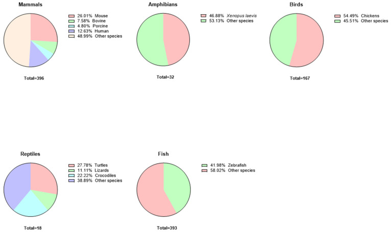
Animal embryos are vital tools in scientific research, providing insights into biological processes and disease mechanisms. This paper explores their historical and contemporary significance, highlighting the shift towards the refinement of in vitro systems as alternatives to animal experimentation. We have conducted a data review of the relevant literature on the use of embryos in research and synthesized the data to highlight the importance of this model for scientific progress and the ethical considerations and regulations surrounding embryo research, emphasizing the importance of minimizing animal suffering while promoting scientific progress through the principles of replacement, reduction, and refinement. Embryos from a wide range of species, including mammals, fish, birds, amphibians, and reptiles, play a crucial experimental role in enabling us to understand factors such as substance toxicity, embryonic development, metabolic pathways, physiological processes, etc., that contribute to the advancement of the biological sciences. To apply this model effectively, it is essential to match the research objectives with the most appropriate methodology, ensuring that the chosen approach is appropriate for the scope of the study.

Keywords: 3Rs; alternatives methods; animal embryos; animal ethics; biomedical research; embryonic development; genetic; model embryos.


Conflict of interest statement

The authors declare no conflicts of interest.

Figures

Figure 1
Figure 1
Phylogenetic tree of vertebrate evolution. This phylogenetic tree illustrates the evolutionary relationships among major vertebrate groups, highlighting key branching events. The diagram shows the emergence of tetrapods from ancestral aquatic vertebrates, marking a transition to terrestrial life. The evolution of amniotes further reflects adaptations to dry environments, leading to the divergence of reptiles, birds, and mammals. These evolutionary nodes represent shared ancestry, emphasizing the conservation of embryonic development, physiological processes, and anatomical features. Understanding these relationships provides a framework for comparative research, aiding studies on developmental biology, evolutionary mechanisms, and the use of model organisms in biomedical research.
Figure 2
Figure 2
Proportional use of vertebrate embryos in research. These pie charts represent the distribution of research utilizing embryos from various vertebrate groups, with species-specific contributions expressed as percentages of the total studies in each group. In mammals, Mus musculus embryos account for 26.01% of research, while 12.63% involve human embryos, with nearly half (48.99%) using fewer common species. Among amphibians, Xenopus laevis makes up 46.88% of studies, reflecting its prominence as a developmental model. Birds are predominantly represented by chickens (54.49%), while zebrafish embryos contribute 41.98% to fish research, underscoring their utility in genetic and developmental studies. Reptile research is more dispersed, with turtles (27.78%) and crocodiles (22.22%) leading, though “other species” account for a significant portion across all groups. These distributions highlight the variable reliance on model organisms, shaping the landscape of embryology and comparative biology.
Figure 3
Figure 3
Pre-implantation embryonic development in mammals. This diagram illustrates the key stages of pre-implantation embryonic development in various mammalian species, categorized into small (A) (rabbits, rats, and mice) and large (B) mammals (cattle, pigs, and humans). The stages depicted range from the initial two-cell embryo to the morula and blastocyst phases. The timeline (in days) provides a comparative view of developmental progression across different species, highlighting variations in the rate and timing of embryonic development. This comparative approach offers insights into the evolutionary adaptations and developmental biology of mammals, contributing to our understanding of embryogenesis and species-specific developmental strategies. These values are averages obtained from the literature. It is important to bear in mind that there are differences among all groups due to species-specific characteristics; however, they remain similar or very close to the values presented.
Figure 3
Figure 3
Pre-implantation embryonic development in mammals. This diagram illustrates the key stages of pre-implantation embryonic development in various mammalian species, categorized into small (A) (rabbits, rats, and mice) and large (B) mammals (cattle, pigs, and humans). The stages depicted range from the initial two-cell embryo to the morula and blastocyst phases. The timeline (in days) provides a comparative view of developmental progression across different species, highlighting variations in the rate and timing of embryonic development. This comparative approach offers insights into the evolutionary adaptations and developmental biology of mammals, contributing to our understanding of embryogenesis and species-specific developmental strategies. These values are averages obtained from the literature. It is important to bear in mind that there are differences among all groups due to species-specific characteristics; however, they remain similar or very close to the values presented.
Figure 4
Figure 4
Methodology for in vitro toxicity testing in mammalian embryos. This diagram illustrates the standard methodology used in mammalian toxicity testing, detailing the process from in vitro fertilization to embryonic development under experimental conditions. The procedure begins with the collection of embryos at the two-cell stage, followed by their incubation in a culture medium containing a toxic substance. The diagram further depicts the progressive stages of in vitro embryonic development during incubation, including cell division and differentiation, leading to the blastocyst stage. This approach is widely utilized in toxicology studies to evaluate the impact of various substances on early embryonic development, providing insights into developmental biology and potential teratogenic effects.
Figure 5
Figure 5
Embryonic development in oviparous animals. This diagram illustrates the key stages of embryonic development in various oviparous species, including amphibians, reptiles, chickens, and zebrafish. The stages depicted range from the initial 2-cell embryo to the morula and blastocyst phases. The timeline (in hours) provides a comparative view of developmental progression across different species, highlighting variations in the rate and timing of embryonic development. This comparative approach offers insights into the evolutionary adaptations and developmental biology of oviparous animals, contributing to our understanding of embryogenesis and species-specific developmental strategies. These values are average and are found in the literature. It is important to bear in mind that there are differences between all the groups, which are due to the specific characteristics of the species but which, however, are similar or very close to the values presented.
Figure 6
Figure 6
Experimental methodology for toxicity studies using chicken embryos. This schematic represents two methodologies employed in using bird embryos, specifically chicken eggs, as models for studying the toxicity of substances. The process begins with the inoculation of a diluted toxic substance on day 0 for the first methodology or the immersion of the egg in a diluted mixture of toxic substance for 30 min. After embryonic development, the eggs are examined for anomalies. This approach allows researchers to assess the impact of toxic substances on embryonic development, providing valuable insights into developmental toxicity and the potential risks associated with exposure to harmful chemicals.
Figure 7
Figure 7
Experimental methodology for toxicity studies using Xenopus laevis embryos. This schematic outlines the common methodology for using amphibian embryos, specifically the Xenopus laevis model, in toxicity studies. The process begins with the collection of oocytes following the superovulation of Xenopus laevis. These oocytes are then subjected to in vitro fertilization. The resulting embryos are exposed to toxins at various stages of development. This method enables researchers to analyze the impact of toxic substances on embryonic growth and differentiation, offering valuable data on the potential risks and effects of environmental toxins on amphibian development.
Figure 8
Figure 8
Experimental methodology for toxicity studies using turtle eggs. This schematic depicts the common methodology for employing turtle eggs as a representative reptile model in ex vivo embryonic toxicity studies. The process involves the collection of fertilized eggs, which are then incubated in an incubator or in an environment with a sandy substrate contaminated with the substance under investigation. This setup allows researchers to study the effects of environmental contaminants on the development of reptile embryos, providing insights into the potential impacts of toxins on embryonic growth and survival in reptilian species.
Figure 9
Figure 9
Experimental methodology for toxicity studies using zebrafish embryos. This schematic illustrates the standard methodology for utilizing zebrafish embryos in toxicity testing and other biomedical experiments. The process begins, normally, with the collection of embryos at the 64-cell stage, approximately 2 h post-fertilization. These embryos are then incubated in a culture medium containing varying concentrations of the toxic substance. Over a 72-h incubation period, embryonic development is closely observed. This method allows researchers to evaluate the impact of toxic substances on early developmental stages, offering critical insights into developmental biology and the potential effects of environmental toxins on aquatic organisms. The zebrafish model is particularly valuable due to its transparency and rapid development, making it an excellent system for studying a wide range of biological processes and toxicological responses.
Figure 10
Figure 10
Embryonic models as alternatives in biomedical experimentation. This illustrative figure highlights the use of embryonic models as viable alternatives in biomedical research, showcasing their advantages, limitations, and diverse applications. Key benefits include the potential to replace, reduce, and refine the use of animals in research. These models are employed in studies ranging from vitro fertilization and reproductive biology to developmental and reproductive toxicology, as well as research on the origins of diseases, mutations, malformations, cell differentiation, and signaling pathways. However, limitations such as the restricted comparability between different embryonic models, ethical and legislative constraints, and issues with reproducibility must be considered. By utilizing these models, researchers can achieve meaningful results without relying on human embryos, thereby addressing ethical concerns while advancing scientific knowledge.

References

    1. Veissier I., Aubert A., Boissy A. Animal welfare: A result of animal background and perception of its environment. Anim. Front. 2012;2:7–15. doi: 10.2527/af.2012-0043. - DOI
    1. Turner P.V., Barbee R.W. Responsible Science and Research Animal Use. Ilar J. 2019;60:1–4. doi: 10.1093/ilar/ilz020. - DOI - PubMed
    1. McCausland C. The Five Freedoms of Animal Welfare are Rights. J. Agric. Environ. Ethics. 2014;27:649–662. doi: 10.1007/s10806-013-9483-6. - DOI
    1. Kim K., Jo W., Lee G.H. Why do we always care about the welfare of laboratory animals? Open Access Gov. 2023;2023:518–519. doi: 10.56367/OAG-36-10808. - DOI
    1. Franco N.H. Animal Experiments in Biomedical Research: A Historical Perspective. Animals. 2013;3:238–273. doi: 10.3390/ani3010238. - DOI - PMC - PubMed

LinkOut - more resources