. 2025 Mar 15;26(6):2659.
doi: 10.3390/ijms26062659.

The Protective Effects of Burdock Fructooligosaccharide on Preterm Labor Through Its Anti-Inflammatory Action

Affiliations

The Protective Effects of Burdock Fructooligosaccharide on Preterm Labor Through Its Anti-Inflammatory Action

Qunfei Ma et al. Int J Mol Sci. .

Abstract

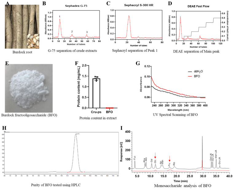
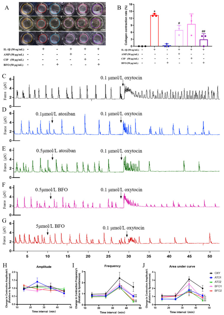
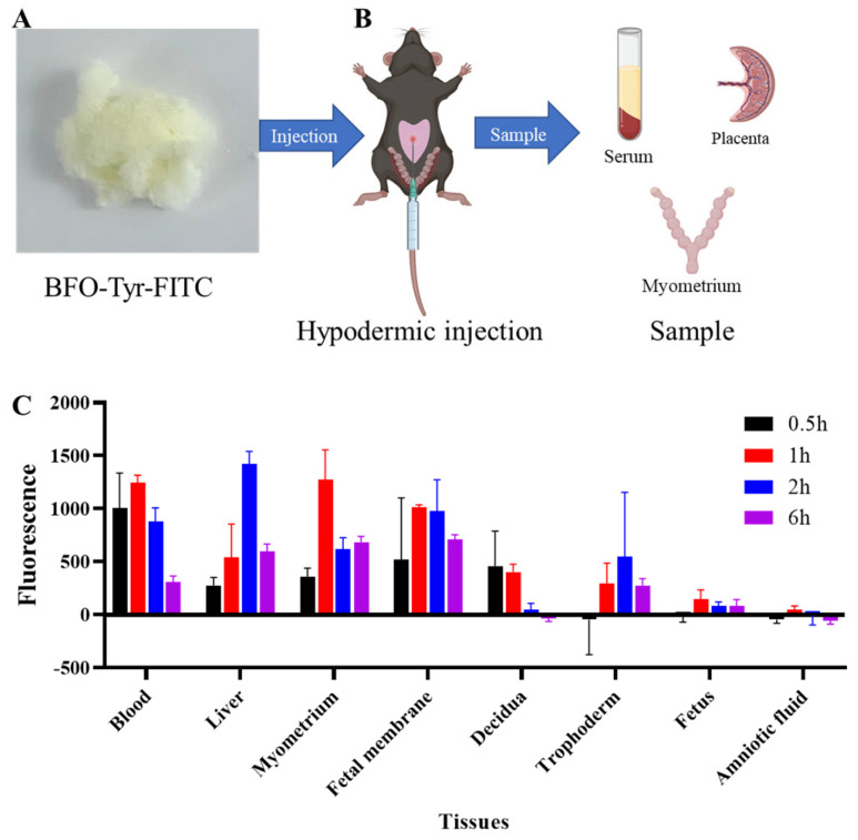
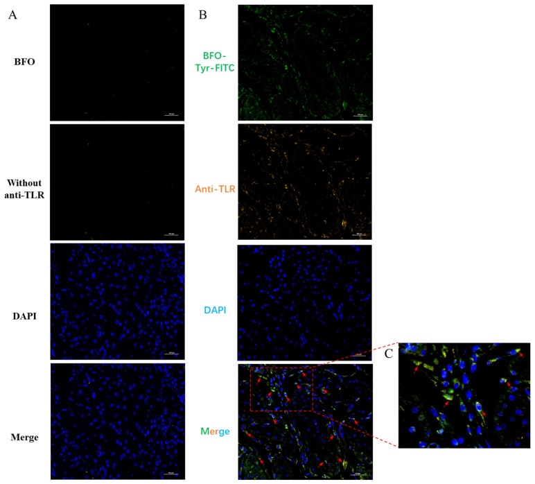
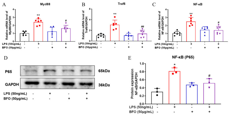
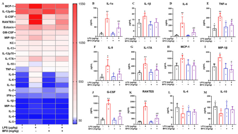
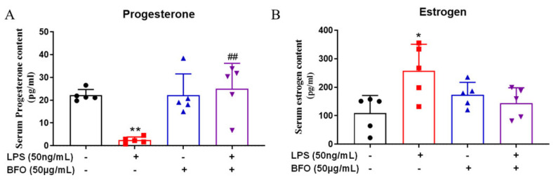
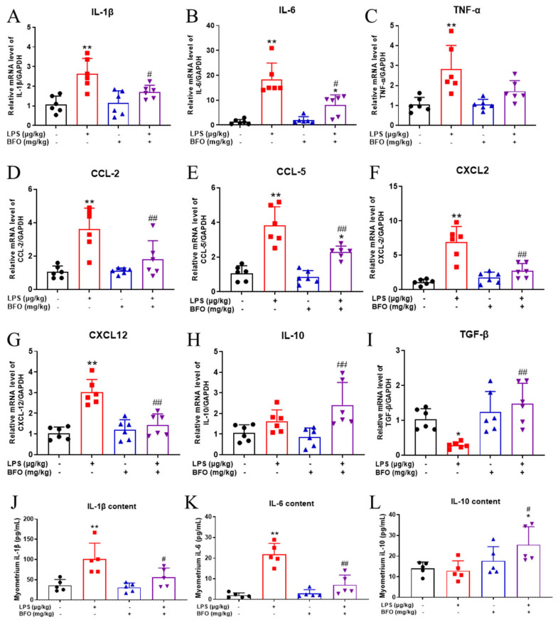
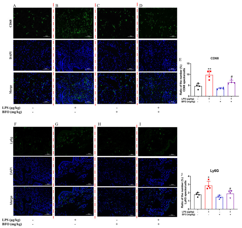
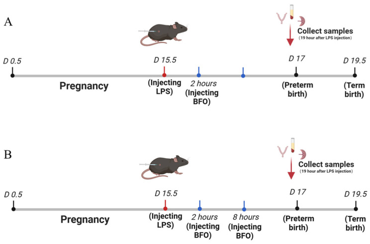
Most pharmacotherapeutic chemicals/interventions used to manage preterm labor (PTL) often cause neonatal morbidity and maternal adverse reactions. Fructooligosaccharides, extracted from traditional Chinese medicine, can alleviate inflammation, demonstrate antiviral capabilities, and protect against antioxidant stress, implying a potential effective PTL treatment. In this study, we explored the protective effects of the purified burdock fructooligosaccharide (BFO), a Gfn-type fructose polymer, on inflammation-induced PTL. It was found that two doses of 30 mg/kg mouse BFO administration to pregnant mice at a 6 h interval can effectively ameliorate lipopolysaccharide (LPS)-induced PTL. Drug dynamic distribution analysis revealed that BFO was rather highly enriched in myometrial tissues, could inhibit oxytocin-induced uterine smooth muscle contraction, and could bind toll-like receptor 4 (TLR4) on the membrane of uterine smooth muscle cells, downregulating the expression of downstream genes, attenuating the upregulation of inflammatory cytokines in serum and the myometrium, as well as reversing the increased macrophage and neutrophil infiltration into the myometrium induced by LPS. It can also interfere with the levels of estrogen and progesterone, alleviating the occurrence of premature birth. These findings collectively suggest that BFO might serve as a promising therapeutic agent for inflammation-related preterm labor to safeguard the health of both the mother and fetus.

Keywords: burdock fructooligosaccharide; inflammation; myometrium; preterm labor; toll-like receptor 4.


Conflict of interest statement

The authors declare no conflicts of interest.

Figures

Figure 1
Figure 1
The preparation and identification of fructooligosaccharide. (A) Fresh burdock root. (B) The elution curve of the separation of crude polysaccharide from burdock root using Sephadex G-75 and followed by further purification of peak 1 utilizing (C) Sephacryl S-300 HR and (D) DEAE-cellulose-52. (E) The lyophilized BFO powder. (F) The protein contents in crude polysaccharide (Cru-ps) and BFO were determined. (G) UV spectral scanning of BFO and distilled water between 230 and 400 nm was conducted. (H) Purity assessment and (I) monosaccharide composition analysis were performed for BFO. In the monosaccharide composition analysis, the black line represents the standard chromatogram, while the red line represents the sample chromatogram, and the red arrows indicate the positions of glucose and fructose.
Figure 2
Figure 2
Determination of myometrial cell contractility and the contractile tension of ex vivo myometrial strips. (A) Collagen contraction of primary uterine smooth muscle cells was determined and (B) their contraction rates were analyzed, the same color circle represents the same treatment replicates. (C) The contraction curve of myometrial strips from pregnant mice was induced by 0.1 μmol/L oxytocin in vitro, and the changes in contractions were induced by oxytocin after preconditioning with different drugs including (D) 0.1 μmol/L atosiban, (E) 0.5 μmol/L atosiban, (F) 0.5 μmol/L BFO, and (G) 5 μmol/L BFO. The relative changes in the contraction (H) amplitude, (I) frequency, and (J) area under the curve were calculated at intervals of every 10 min compared with the 10 min spontaneous contractions before the administration of the different reagents. * p ≤ 0.05 and ** p ≤ 0.01 vs. control group; # p ≤ 0.05 and ## p ≤ 0.01 vs. LPS group using one-way ANOVA with Tukey’s HSD test. N = 3.
Figure 3
Figure 3
Drug distribution kinetics of BFO in vivo. (A) The dry powder of BFO-Tyr-FITC. (B) The schematic diagram of BFO-Tyr-FITC injection and tissue collection. (C) Histogram of fluorescence intensity changes in the serum, liver, myometrium, fetal membrane, decidua, trophoderm, fetus, and amniotic fluid at 0.5, 1, 2, and 6 h after injection, respectively.
Figure 4
Figure 4
Co-localization of BFO and TLR4 on myometrial cells. The immunofluorescence co-localization between BFO (green) and TLR4 (orange) in mouse myometrium in vivo was determined for (A) the control group (without fluorescein) and (B) the treatment group (labeled with fluorescein), along with its locally magnified images (C). The bright yellow colors pointed by red arrows indicate the co-localization of BFO with TLR4.
Figure 5
Figure 5
Inhibitory effects of BFO on key downstream genes of TLR4 signaling pathway in the myometrium. The mRNA expression of MyD88 (A), Traf6 (B), and NF-κB (C) genes, as well as the protein expression of NF-κB subunit P65 (D) and its statistical analysis (E) in USMCs were presented. The cells were treated with BFO (50 μg/mL) and LPS (50 ng/mL) alone or in combination for 24 h. * p ≤ 0.05 and ** p ≤ 0.01 vs. control group; # p ≤ 0.05 and ## p ≤ 0.01 vs. LPS group using one-way ANOVA with Tukey’s HSD test. N ≥ 3.
Figure 6
Figure 6
Inflammatory cytokine levels in maternal serum were analyzed using Luminex technology. (A) Heat map results of inflammatory cytokines in the sera of pregnant mice were obtained after 19 h of LPS and/or BFO injection. Significant differences were observed in pro-inflammatory and chemotactic cytokines, including (B) IL-1α, (C) IL-1β, (D) IL-6, (E) IL-9, (F) IL-12p70, (G) IL-17A, (H) MCP-1, (I) MIP-1β, (J) G-CSF, and (K) RANTES, as well as levels of anti-inflammatory cytokines (L) IL-4 (M) and IL-10 among different groups. * p ≤ 0.05 and ** p ≤ 0.01 vs. control group; # p ≤ 0.05 and ## p ≤ 0.01 vs. LPS group using one-way ANOVA with Tukey’s HSD test. N = 4.
Figure 7
Figure 7
Progesterone and estradiol levels in maternal serum were analyzed using ELISA kits. The changes in the (A) progesterone content and (B) estradiol content in the sera of pregnant mice were obtained after 19 h of LPS and/or BFO injection. * p ≤ 0.05; ** p ≤ 0.01 vs. control group; ## p ≤ 0.01 vs. LPS group using one-way ANOVA with Tukey’s HSD test. N = 4.
Figure 8
Figure 8
The mRNA and protein expressions of inflammatory cytokines in the myometrium. The mRNA expression levels of inflammation-related cytokines in the myometrium, including (A) IL-1β, (B) IL-6, (C) TNF-α, (D) CCL-2, (E) CCL-5, (F) CXCL2, (G) CXCL12, (H) IL-10, and (I) TGF-β were examined. And changes in the content of inflammatory cytokines, including (J) IL-1β, (K) IL-6, and (L) IL-10, in the myometrium were also analyzed. * p ≤ 0.05 and ** p ≤ 0.01 vs. control group; # p ≤ 0.05 and ##p ≤ 0.01 vs. LPS group using one-way ANOVA with Tukey’s HSD test. N ≥ 5.
Figure 9
Figure 9
The infiltration of inflammatory cells into the myometrium. Macrophage infiltration was photographed at 40× magnification in the (A) control group, (B) LPS administration group, (C) double dose of BFO administration group, and (D) LPS combined with double doses of BFO administration group; (E) their positive staining rate statistics. Neutrophil infiltration was similarly imaged at 40× magnification in the (F) control group, (G) LPS administration group, (H) double dose of BFO administration group, and (I) LPS combined with double doses of BFO administration group; (J) their positive staining rate statistics. Red dashed lines are used to distinguish immunofluorescence staining for different treatments, with green representing target protein fluorescence and blue representing DAPI staining. Scale bar: 100 μm. * p ≤ 0.05 and ** p ≤ 0.01 vs. control group; # p ≤ 0.05 vs. LPS group using one-way ANOVA with Tukey’s HSD test. N = 4.
Figure 10
Figure 10
The scheme of establishing an animal model for preterm labor and the single injection (A) and double injection (B) of BFO treatment.

Similar articles

References

    1. Sharrow D., Hug L., You D., Alkema L., Black R., Cousens S., Croft T., Gaigbe-Togbe V., Gerland P., Guillot M., et al. Global, regional, and national trends in under-5 mortality between 1990 and 2019 with scenario-based projections until 2030: A systematic analysis by the UN Inter-agency Group for Child Mortality Estimation. Lancet Glob. Health. 2022;10:e195–e206. doi: 10.1016/S2214-109X(21)00515-5. - DOI - PMC - PubMed
    1. Ward V.C., Lee A.C.C., Hawken S., Otieno N.A., Mujuru H.A., Chimhini G., Wilson K., Darmstadt G.L. Overview of the Global and US Burden of Preterm Birth. Clin. Perinatol. 2024;51:301–311. doi: 10.1016/j.clp.2024.02.015. - DOI - PubMed
    1. Gomez-Lopez N., Galaz J., Miller D., Farias-Jofre M., Liu Z., Arenas-Hernandez M., Garcia-Flores V., Shaffer Z., Greenberg J.M., Theis K.R., et al. The immunobiology of preterm labor and birth: Intra-amniotic inflammation or breakdown of maternal–fetal homeostasis. Reproduction. 2022;164:R11–R45. doi: 10.1530/REP-22-0046. - DOI - PMC - PubMed
    1. Taylor J., Sharp A., Rannard S.P., Arrowsmith S., McDonald T.O. Nanomedicine strategies to improve therapeutic agents for the prevention and treatment of preterm birth and future directions. Nanoscale Adv. 2023;5:1870–1889. doi: 10.1039/D2NA00834C. - DOI - PMC - PubMed
    1. Miller F.A., Sacco A., David A.L., Boyle A.K. Interventions for Infection and Inflammation-Induced Preterm Birth: A Preclinical Systematic Review. Reprod. Sci. 2023;30:361–379. doi: 10.1007/s43032-022-00934-x. - DOI - PMC - PubMed

MeSH terms

LinkOut - more resources