. 2025 Mar 16;26(6):2668.
doi: 10.3390/ijms26062668.

Plasma Exosomal-Derived SERPINA1 and GNAI2 Downregulation as Potential Diagnostic Biomarkers of Kawasaki Disease with Coronary Artery Aneurysms

Affiliations

Plasma Exosomal-Derived SERPINA1 and GNAI2 Downregulation as Potential Diagnostic Biomarkers of Kawasaki Disease with Coronary Artery Aneurysms

Yang Zheng et al. Int J Mol Sci. .

Abstract

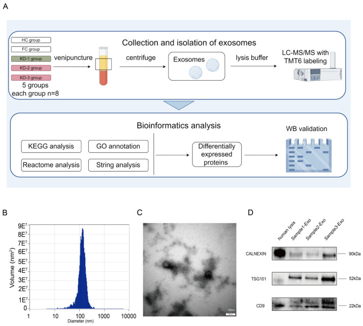
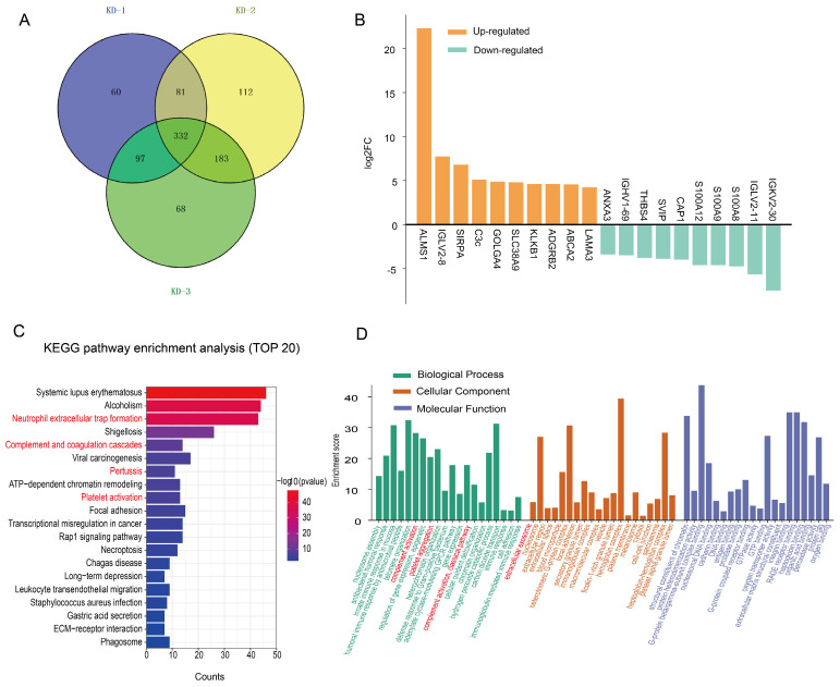
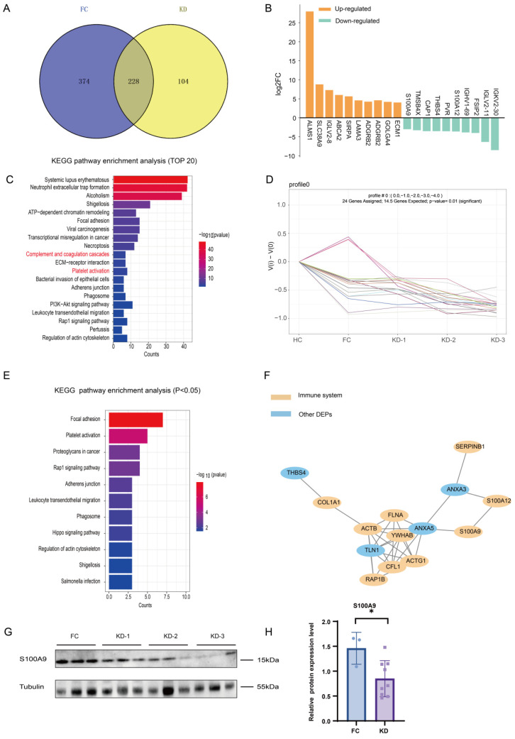
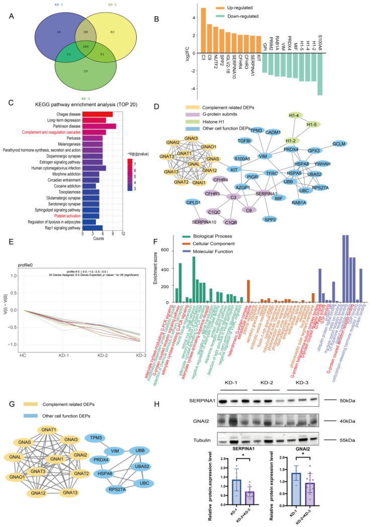
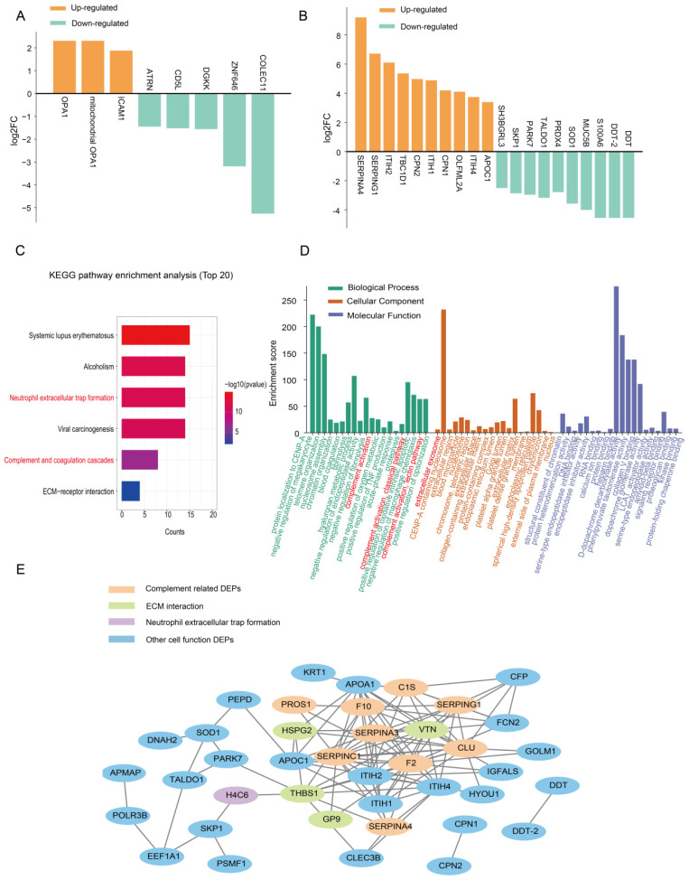
Kawasaki disease (KD) with coronary artery aneurysms (CAAs) is currently the primary cause of childhood acquired heart disease with an unclear pathogenesis. We established five groups for the discovery of differentially expressed proteins (DEPs): healthy control, febrile control, KD without CAAs, KD with small and medium CAAs, and KD with giant CAAs (n = 8 in each group). The validation of selected DEPs was conducted in another five groups (n = 4 in each group). We conducted comprehensive bioinformatics analyses to elucidate the functional roles of the DEPs in the groups of KD with CAAs and KD without CAAs. A total of 104 DEPs were identified in KD patients, which were primarily associated with complement-related pathways. A trend analysis of these 104 DEPs revealed 54 significantly changed DEPs associated with increased disease severity, which were primarily associated with G-protein-related functions. The alterations in α-1-antitrypsin short peptide (SERPINA1) and guanine nucleotide-binding protein G(i) subunit alpha-2 (GNAI2), which were selected from complement-related and G-protein-related pathways, respectively, were validated by Western blotting, and they were significantly decreased in KD patients with vs. without CAAs. In addition, we conducted an analysis of the DEPs in the groups of KD with CAAs and KD without CAAs, separately. There were 91 DEPs specifically expressed in KD patients with CAAs, associated with the neutrophil extracellular trap and complement pathways, while 16 DEPs were specific to those without CAAs, associated with viral infection and immunity pathways. Additionally, for DEPs among different severities of CAAs, there were 102 DEPs in KD patients with small and medium CAAs, associated with complement pathways and platelet activation pathways, whereas 34 DEPs were specific to giant CAAs, associated with the Rap1 signaling pathway and cell functions. In conclusion, this study provides plasmatic exosomal protein profiles in KD patients with CAAs, suggesting that SERPINA1 and GNIA2 might serve as novel potential diagnostic biomarkers for KD with CAAs.

Keywords: Kawasaki disease (KD); coronary artery aneurysms (CAAs); exosome; proteomics.


Conflict of interest statement

The authors declare no conflicts of interest.

Figures

Figure 1
Figure 1
Flowchart and confirmation of plasma exosomes. (A) Flow diagrams of study design, classification of recruited children, and bioinformatics analysis workflow (image created with Figdraw 2.0, https://www.figdraw.com/static/index.html#/ and published with permission). KD-1: KD patients without CAAs, KD-2: KD patients with small and medium CAAs, KD-3: KD patients with giant CAAs. KD patients without and with CAAs are highlighted in green and pink, respectively. (B) Characterization by NTA of exosomes isolated from plasma of subjects, showing centralized range of diameters. (C) Transmission electron microscopy image of exosomes isolated from plasma of subjects. (D) Western blot of exosome protein markers (exosome markers CD9 and TSG101 and cell organelle marker CALNEXIN) in healthy children plasma and isolated exosomes. Equal amounts of protein were loaded in each lane.
Figure 2
Figure 2
Protein expression profiles of plasma exosomes in KD patients compared with the HC group. (A) Venn diagram showing the number of DEPs in KD patients; 332 overlapped exosomal DEPs were commonly expressed among three subgroups. (B) The top 10 up-/downregulated proteins in the KD patients. (C) KEGG pathway enrichment analysis of 332 exosomal DEPs expressed in KD patients, showing the top 20 enriched pathways. (D) GO annotations for 332 exosomal DEPs expressed in KD patients. The red font referred to the pathways we focused and mentioned.
Figure 3
Figure 3
Protein expression profiles of plasma exosomes expressed in KD patients compared with the FC group. (A) Venn diagram showing the number of DEPs in the KD group compared with the FC group. (B) The top 10 up-/downregulated proteins both in KD patients and in the FC group. (C) KEGG pathway enrichment analysis of 228 expressed exosomal DEPs in the KD group, showing the top 20 enriched pathways. (D) Line graph showing expression changes in 24 exosomal DEPs with downregulated expression. (E) KEGG pathway enrichment analysis of 24 downregulated exosomal DEPs in Profile 0, showing all the enriched pathways with significant differences (p < 0.05). (F) STRING analysis of the PPI network among the 24 exosomal DEPs. (G) WB analysis to validate the expression level of S100A9 in KD patients and the FC group, revealing significant differences in KD patients compared to the HC group. The expression levels were normalized to α-Tubulin. (H) A bar chart showing the expression level of S100A9 in KD patients and the FC group. * indicates p < 0.05. The red font referred to the pathways we focused and mentioned.
Figure 4
Figure 4
Protein expression profiles of plasma exosomes expressed in the KD groups without and with CAAs. (A) A Venn diagram showing the number of DEPs in the KD-1, KD-2, KD-3, and FC groups. (B) The top 10 up-/downregulated proteins in KD patients without and with CAAs. (C) KEGG pathway enrichment analysis of 104 expressed exosomal DEPs in KD patients, showing the top 20 enriched pathways. (D) STRING analysis of the PPI network among the 104 exosomal DEPs. (E) A line graph showing the expression changes in 54 exosomal DEPs with downregulated expression in Profile 0. (F) GO annotations for 54 exosomal DEPs in Profile 0. (G) STRING analysis of the PPT network for the 54 exosomal DEPs. (H) WB analysis of SERPINA1 and GNAI2 in the KD groups without and with CAAs; 2 DEPs revealed significant differences. The expression levels were normalized to α-Tubulin. The bar chart shows the expression level. * indicates p < 0.05. KD-1 group: KD patients without CAAs; KD-2 + KD-3 groups: KD patients with CAAs. The red font referred to the pathways we focused and mentioned.
Figure 5
Figure 5
Protein expression profiles of plasma exosomes in the KD groups without and with CAAs separately. (A) The top up-/downregulated proteins in KD patients without CAAs, showing all upregulated DEPs and the top five downregulated DEPs. (B) The top 10 up-/downregulated proteins in KD patients with CAAs. (C) KEGG pathway enrichment analysis of 91 exosomal DEPs expressed in the KD-2 + KD-3 groups, showing all enriched pathways. (D) GO annotations for 91 exosomal DEPs expressed in the KD-2 + KD-3 groups. (E) STRING analysis of the PPT network among the 91 exosomal DEPs. The red font referred to the pathways we focused and mentioned.

Similar articles

References

    1. McCrindle B.W., Rowley A.H., Newburger J.W., Burns J.C., Bolger A.F., Gewitz M., Baker A.L., Jackson M.A., Takahashi M., Shah P.B., et al. Diagnosis, treatment, and long-term management of kawasaki disease: A scientific statement for health professionals from the American Heart Association. Circulation. 2017;27:e927–e999. doi: 10.1161/CIR.0000000000000484. - DOI - PubMed
    1. Burns J.C., Glodé M.P. Kawasaki syndrome. Lancet. 2004;364:533–544. doi: 10.1016/S0140-6736(04)16814-1. - DOI - PubMed
    1. Newburger J.W., Takahashi M., Burns J.C. Kawasaki Disease. J. Am. Coll. Cardiol. 2016;67:1738–1749. doi: 10.1016/j.jacc.2015.12.073. - DOI - PubMed
    1. Goel A.R., Yalcindag A. An Update on Kawasaki Disease. Curr. Rheumatol. Rep. 2024;27:4. doi: 10.1007/s11926-024-01167-4. - DOI - PubMed
    1. Wu M.H., Chen H.C., Yeh S.J., Lin M.T., Huang S.C., Huang S.K. Prevalence and the long-term coronary risks of patients with kawasaki disease in a general population <40 years. Circ. Cardiovasc. Qual. Outcomes. 2012;5:566–570. - PubMed

MeSH terms