. 2025 Mar 16;26(6):2673.
doi: 10.3390/ijms26062673.

Developmental Endothelial Locus-1 Promotes Osteoclast Differentiation and Activation

Affiliations

Developmental Endothelial Locus-1 Promotes Osteoclast Differentiation and Activation

Kentaro Imamura et al. Int J Mol Sci. .

Abstract

Developmental endothelial locus-1 (DEL-1) has traditionally been characterized within the scientific community as having anti-inflammatory properties with potential inhibitory effects on osteoclast formation. Our investigation challenges this paradigm by examining Del-1 expression in RAW264.7 cells and bone marrow-derived macrophages (BMMs) during osteoclastogenesis, as well as its functional impact on osteoclast development and activity. Our experimental findings revealed that Del-1 mRNA levels were markedly elevated in cells stimulated by the receptor activator of the nuclear factor κB ligand compared to unstimulated precursors. When cultured with varying concentrations of recombinant DEL-1, osteoclast differentiation increased in a dose-dependent manner. Furthermore, BMMs isolated from ovariectomized mice exhibited significantly higher Del-1 mRNA expression than those from control animals. To confirm DEL-1's role, we employed RNA interference techniques, demonstrating that DEL-1 silencing in RAW264.7 cells substantially reduced osteoclast formation. These results suggest that DEL-1 plays a previously unrecognized role in promoting osteoclastogenesis and may contribute to bone metabolism imbalances in conditions like osteoporosis, highlighting its complex role in skeletal homeostasis and its potential as a therapeutic target.

Keywords: DEL-1; bone metabolism; osteoclasts; osteoporosis.


Conflict of interest statement

Authors Keita Tachi, Hironori Kasai, and Ryutaro Shohara private practitioners and each one works at a private office. The remaining authors declare that the research was conducted in the absence of any commercial or financial relationships that could be construed as a potential conflict of interest.

Figures

Figure 1
Figure 1
Del-1 mRNA expression and cellular viability in RAW 264.7 cells and BMMs. After RAW 264.7 cells (ac) and BMMs (df) were cultured in the presence of the receptor activator of nuclear factor κB ligand (RANKL), Del-1 mRNA was measured and cell viability was assessed. The significant upregulation of Del-1 mRNA expression in both RAW 264.7 cells (a) and BMMs (d) is observed in RANKL-activated cells compared to preosteoclasts. Cell viability assessment using trypan blue (b,e) and MTT assays (c,f) at various DEL-1 concentrations (0.1–1000 ng/mL). Differential cellular responses are shown between RAW264.7 cells and BMMs, highlighting concentration-dependent effects. Data are expressed as mean ± standard deviation (SD) (n = 9). * p < 0.05, ** p < 0.01, and *** p < 0.001.
Figure 2
Figure 2
The effect of DEL-1 on osteoclast differentiation. TRAP staining images demonstrating osteoclast formation in response to DEL-1 treatment. RAW264.7 cells (a) and BMMs (b) treated with RANKL and varying concentrations of DEL-1 (0.1–10 ng/mL). The numbers of TRAP-positive cells containing more than 3 nuclei were counted as osteoclasts. Data are expressed as mean ± SD (n = 9). * p < 0.05, ** p < 0.01, and *** p < 0.001.
Figure 3
Figure 3
Osteoclast differentiation marker gene expression. Real-time RT-PCR analysis of osteoclast differentiation marker genes in RAW264.7 cells (a) and BMMs (b). Expression levels of carbonic anhydrase II (CaII), colony-stimulating factor receptor (C-fms), matrix metalloproteinase-9 (Mmp-9), and cathepsin K (Cat-K) in RAW264.7 cells and BMMs under various DEL-1 concentrations. Significant upregulation of differentiation markers is observed, indicating comprehensive molecular response to DEL-1 treatment. Data are expressed as mean ± SD (n = 9). * p < 0.05, ** p < 0.01, and *** p < 0.001.
Figure 4
Figure 4
The effect of DEL-1 on osteoclast activation. Pit resorption assay demonstrating enhanced bone resorption capabilities. Pit area images showing expanded resorption areas in DEL-1 treated groups (a). Quantification of pit areas using ImageJ (Version 1.52) (b). Comparative analysis demonstrates the functional implications of DEL-1 treatment on osteoclast bone resorption capabilities. Original magnification × 10, bars = 100 μm. Data are expressed as mean ± SD (n = 6). * p < 0.05, ** p < 0.01, and *** p < 0.001.
Figure 5
Figure 5
Ovariectomized (OVX) mouse model. Micro-computed tomography (µCT) imaging of bone metabolism changes. Representative µCT images showing structural alterations in OVX mice compared to sham-operated controls (Control) (a). Bars = 500 μm. Quantitative analysis of bone volume fractions, trabecular separation, and structure model index (b). Abbreviations indicate bone volume (BV), total volume (TV), structure model index (SMI), trabecular separation (Tb. Sp), and trabecular number (Tb.N). The mRNA expression of Del-1, Rank, Cat-K, and Mmp-9 was measured in BMMs collected from mice with OVX and/or RANKL by real-time RT-PCR (c). Molecular profiling of BMMs demonstrating elevated Del-1 mRNA expression and upregulation of osteoclast differentiation markers in OVX mice. Data are expressed as mean ± SD (n = 5). * p < 0.05, ** p < 0.01, and *** p < 0.001.
Figure 6
Figure 6
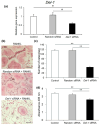
Del-1 siRNA suppressed osteoclast differentiation. Small interfering RNA (siRNA) experiments revealed the functional significance of DEL-1 in RAW264.7 cells. Confirmation of Del-1 mRNA expression reduction using siRNA (a). TRAP staining images showing the effect of Del-1 siRNA on osteoclast differentiation (b). Original magnification × 10, bars = 100 μm. The numbers of TRAP-positive cells containing more than 3 nuclei were counted as osteoclasts (c). Quantification of TRAP-positive multinucleated cells demonstrating 38% reduction in osteoclast differentiation. Osteoclast formation was measured by a TRAP-solution assay (d). Statistical analysis of Del-1 mRNA expression and osteoclast differentiation following siRNA treatment (d). Data are expressed as mean ± SD (n = 9). * p < 0.05 and ** p < 0.01.
Figure 7
Figure 7
Proposed mechanism of bone resorption regulation by DEL-1. This schematic diagram illustrates the molecular pathways through which DEL-1 promotes osteoclast differentiation and activation. The left panel depicts how DEL-1 interacts on osteoclast precursors, activating intracellular signaling cascades via RANK-RANKL interaction. These signals upregulate the expression of osteoclast-specific genes (Cat-K, MMP-9, etc.) and promote the cytoskeletal reorganization necessary for mature osteoclast formation. The right panel illustrates how estrogen deficiency alters this pathway, showing increased Del-1 expression and enhanced RANKL signaling, collectively promoting excessive osteoclastogenesis and bone resorption. Red arrows indicate upregulation.

Similar articles

References

    1. Vernal R., Dutzan N., Chaparro A., Puente J., Valenzuela M.A., Gamonal J. Levels of interleukin-17 in gingival crevicular fluid and in supernatants of cellular cultures of gingival tissue from patients with chronic periodontitis. J. Clin. Periodontol. 2005;32:383–389. doi: 10.1111/j.1600-051X.2005.00684.x. - DOI - PubMed
    1. Li J., Chen X., Lu L., Yu X. The relationship between bone marrow adipose tissue and bone metabolism in postmenopausal osteoporosis. Cytokine Growth Factor Rev. 2020;52:88–98. doi: 10.1016/j.cytogfr.2020.02.003. - DOI - PubMed
    1. Takayanagi H. Inflammatory bone destruction and osteoimmunology. J. Periodontal Res. 2005;40:287–293. doi: 10.1111/j.1600-0765.2005.00814.x. - DOI - PubMed
    1. Walsh M.C., Kim N., Kadono Y., Rho J., Lee S.Y., Lorenzo J., Choi Y. Osteoimmunology: Interplay between the immune system and bone metabolism. Annu. Rev. Immunol. 2006;24:33–63. doi: 10.1146/annurev.immunol.24.021605.090646. - DOI - PubMed
    1. Rodan G.A., Martin T.J. Therapeutic approaches to bone diseases. Science. 2000;289:1508–1514. doi: 10.1126/science.289.5484.1508. - DOI - PubMed

LinkOut - more resources