. 2025 Mar 17;26(6):2705.
doi: 10.3390/ijms26062705.

Activin A Inhibitory Peptides Suppress Fibrotic Pathways by Targeting Epithelial-Mesenchymal Transition and Fibroblast-Myofibroblast Transformation in Idiopathic Pulmonary Fibrosis

Affiliations

Activin A Inhibitory Peptides Suppress Fibrotic Pathways by Targeting Epithelial-Mesenchymal Transition and Fibroblast-Myofibroblast Transformation in Idiopathic Pulmonary Fibrosis

Victor Alexandre F Bastos et al. Int J Mol Sci. .

Abstract

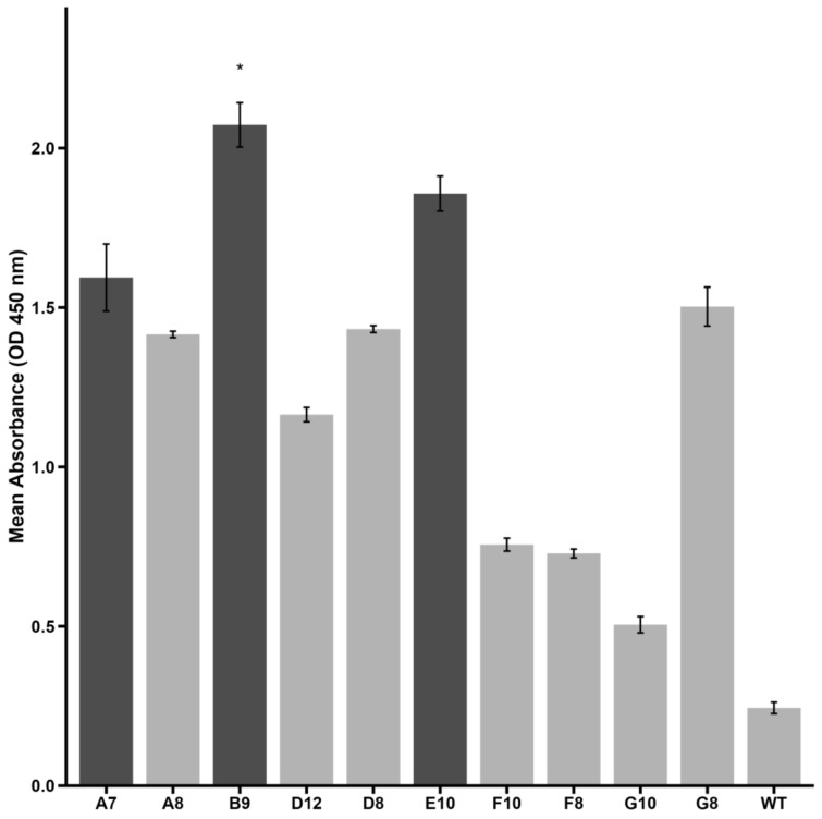
Idiopathic pulmonary fibrosis (IPF) is a progressive and incurable chronic interstitial lung disease characterized by excessive fibrosis and impaired lung function. Current treatments, such as pirfenidone and nintedanib, slow disease progression but fail to halt or reverse fibrosis, highlighting the need for novel approaches. Activin A, which belongs to the TGF-β superfamily, is implicated in various fibrosis-related mechanisms, including epithelial-mesenchymal transition (EMT), a process where epithelial cells acquire mesenchymal characteristics, and fibroblast-myofibroblast transformation (FMT), in which fibroblasts differentiate into contractile myofibroblasts. It also promotes inflammatory cytokine release and extracellular matrix buildup. This study aimed to inhibit Activin A activity using synthetic peptides identified through phage display screening. Of the ten peptides isolated, A7, B9, and E10 demonstrated high binding affinity and inhibitory activity. Computational modeling confirmed that these peptides target the receptor-binding domain of Activin A, with peptide E10 exhibiting superior efficacy. Functional assays showed that E10 reduced cell migration, inhibited EMT in A549 cells, and suppressed FMT in fibroblast cultures, even under pro-fibrotic stimulation with TGF-β. These findings underscore the therapeutic potential of targeting Activin A with synthetic peptides, offering a promising avenue for IPF treatment and expanding the arsenal of anti-fibrotic strategies.

Keywords: Activin A; anti-fibrotic therapy; epithelial–mesenchymal transition; fibroblast–myofibroblast transformation; idiopathic pulmonary fibrosis; synthetic peptides.


Conflict of interest statement

The authors state that they have no conflicts of interest. The funding sources played no part in the study’s design, data collection, analysis, or interpretation, nor in the writing of the manuscript or the decision to publish the findings.

Figures

Figure 1
Figure 1
Binding affinity of selected phage clones to Activin A as measured by phage–ELISA. Optical density (OD) values at 450 nm are shown for each peptide. Clone B9 demonstrated significantly higher reactivity compared to wild-type phage (negative control) (* p < 0.05, Kruskal–Wallis test with Dunn’s post hoc). Clones A7 and E10 presented an ELISA index > 2 and are also highlighted. Darker bars represent high-binding peptides selected for further investigation. Data are presented as mean ± SE from triplicate experiments.
Figure 2
Figure 2
Structural predictions of molecules and interactions analyzed in this study. (A) Dimeric structure of Activin A (PDB: 2ARV). (B) Interaction of Activin A (gray) with its receptor ActRIIB (yellow). (C) Interaction of Activin A (gray) with follistatin (purple). (DF) Structural predictions of peptides A7, B9, and E10, respectively. (GI) Predicted interactions of Activin A (gray) with peptides A7, B9, and E10, respectively. The conserved region corresponding to the pIII protein sequence of M13 phage is shown in green for all peptides.
Figure 3
Figure 3
Binding interactions between peptides A7, B9, and E10 and the Activin A molecule. (A) The pIII protein region of peptide A7 interacts with isoleucine (I206), lysine (K208), and aspartic acid (D210) within the ActRIIB recognition site of Activin A. (B) Peptide B9 forms a covalent interaction exclusively with lysine (K208) in the ActRIIB recognition site. (C) Peptide E10 interacts with isoleucine (I206), lysine (K208), and aspartic acid (D210) in the ActRIIB recognition site, with a specific contribution from proline in its sequence, enhancing its binding stability. The conserved pIII region is shown in green, Activin A in gray, and the unique peptide regions in cyan (A7), purple (B9), and orange (E10).
Figure 4
Figure 4
Cytotoxic effects of peptides A7, B9, and E10 on A549 cells. Cells were treated with peptides at concentrations of 1 µM, 10 µM, and 50 µM in the presence or absence of pm26TGF-β1 (1 ng/mL) as positive and negative controls, respectively. Cell viability was monitored at 24, 48, and 72 h. Significant cytotoxicity was observed for all peptides at 50 µM at 48 h. After 72 h, only peptides A7 and B9 presented significant cytotoxicity at 50 µM in the presence of pm26TGF-β1. Statistical analysis was conducted using pairwise t-tests with p-values adjusted by the False Discovery Rate (FDR) method (* adjusted p < 0.05, ** adjusted p < 0.01, *** adjusted p < 0.001). Data are presented as mean ± SD from triplicate experiments.
Figure 5
Figure 5
Effects of peptides A7, B9, and E10 on cellular migration. Scratch assays were performed using A549 cells treated with peptides at 1 µM, 10 µM, and 50 µM, in the presence or absence of pm26TGF-β1 (1 ng/mL). Control cells were treated with DMEM medium only in the − pm26TGF-β group and DMEM medium supplemented with pm26TGF-β1 (1 ng/mL) in the + pm26TGF-β1 group. Wound closure was monitored at 24 and 48 h. Peptide E10 exhibited the most consistent and significant inhibitory effects on cellular migration across conditions, particularly at moderate concentrations and in the presence or absence of pm26TGF-β1. Statistical analysis was conducted using pairwise t-tests with p-values adjusted by the False Discovery Rate (FDR) method (* adjusted p < 0.05, ** adjusted p < 0.01, *** adjusted p < 0.001). Data are presented as mean ± SD from triplicate experiments.
Figure 6
Figure 6
Effects of peptide E10 on epithelial–mesenchymal transition (EMT) in A549 cells. Cells were treated with E10 at 1 µM, 10 µM, and 50 µM in the presence or absence of pm26TGF-β1 (1 ng/mL), as positive and negative controls, respectively. EMT inhibition was evaluated by measuring fibronectin expression. The strongest inhibitory effects were observed at 10 µM, with reduced fibronectin levels compared to cells treated only with pm26TGF-β1. DAPI (4′,6-Diamidino-2-phenylindole) was used for nucleus visualization. Scale bar: 200 µm.
Figure 7
Figure 7
Effects of peptide E10 on fibroblast–myofibroblast transformation (FMT) in primary fibroblasts. Cells were treated with E10 at 1 µM, 10 µM, and 50 µM in the presence or absence of pm26TGF-β1 (1 ng/mL), as positive and negative controls, respectively. FMT inhibition was assessed by measuring α-smooth muscle actin (α-SMA) fiber expression. Significant inhibition of FMT was observed at 10 µM and 50 µM, with reduced α-SMA levels compared to cells treated only with pm26TGF-β1. DAPI (4′,6-Diamidino-2-phenylindole) was used for nucleus visualization. Scale bar: 200 µm.

Similar articles

References

    1. Urteaga M.B., Ruz J.R., González M.S. Idiopathic Pulmonary Fibrosis. Radiol. (Engl. Ed.) 2022;64:227–239. doi: 10.1016/j.rxeng.2022.10.009. - DOI - PubMed
    1. Spagnolo P., Kropski J.A., Jones M.G., Lee J.S., Rossi G., Karampitsakos T., Maher T.M., Tzouvelekis A., Ryerson C.J. Idiopathic Pulmonary Fibrosis: Disease Mechanisms and Drug Development. Pharmacol. Ther. 2021;222:107798. doi: 10.1016/j.pharmthera.2020.107798. - DOI - PMC - PubMed
    1. George P.M., Spagnolo P., Kreuter M., Altinisik G., Bonifazi M., Martinez F.J., Molyneaux P.L., Renzoni E.A., Richeldi L., Tomassetti S., et al. Progressive Fibrosing Interstitial Lung Disease: Clinical Uncertainties, Consensus Recommendations, and Research Priorities. Lancet Respir. Med. 2020;8:925–934. doi: 10.1016/S2213-2600(20)30355-6. - DOI - PubMed
    1. Koudstaal T., Wijsenbeek M.S. Idiopathic Pulmonary Fibrosis. Presse Medicale. 2023;52:104166. doi: 10.1016/j.lpm.2023.104166. - DOI - PubMed
    1. Glass D.S., Grossfeld D., Renna H.A., Agarwala P., Spiegler P., DeLeon J., Reiss A.B. Idiopathic Pulmonary Fibrosis: Current and Future Treatment. Clin. Respir. J. 2022;16:84–96. doi: 10.1111/crj.13466. - DOI - PMC - PubMed

MeSH terms

LinkOut - more resources